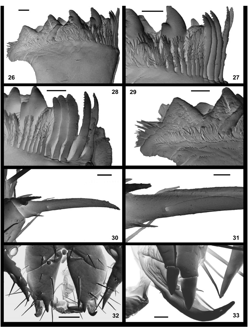
Description: Length (anterior margin of head to posterior margin of telson) up to 5.0 mm; length of head shield up to 0.6 mm. Colour (in absolute ethanol): Head shield, maxillipedes, and posterior trunk tergites and sternites pale orange; anterior trunk tergites and sternites yellow; some specimens uniformly medium orange on head and along length of trunk; legs yellow. Head: Transverse suture extends back to more than 35 % length of head shield. Median notch strong; longitudinal median furrow deep in anterior twothirds, lightly impressed to transverse suture. Region distal to antennocellar sutures depigmented but not entirely so. Setae on head shield symmetrically arranged, four pairs between antennocellar and transverse suture, nine pairs behind these sutures (Fig. 8). Antenna 2.2 2.6 times length of head shield; 15 articles, basal two enlarged, next 12 each about as long as wide, submoniliform, with numerous trichoid sensilla arranged in three indistinctly defined rows (Fig. 13); distal article about twice length of penultimate, bearing two sickleshaped thinwalled basiconic sensilla (Figs. 14, 15); cluster of about eight sensilla brachyconica at tip of distal article (Fig. 14); other articles with a single clavate thinwalled basiconic sensillum on dorsolateral anterior edge of article (Fig. 13), single minute thickwalled basiconic sensillum on mediolateral anterior edge; several sensilla microtrichoidea on proximal part of first article on its dorsal side. Tömösváry organ large, longitudinally ovate, outer margin at lateral edge of cephalic pleurite (Fig. 12). Clypeal apex with cluster of seven setae, three along margins, one medially; transverse band of four clypeal setae in front of labrum; transverse seta projects from pit in sidepiece (Fig. 10). Labral margin prominently incised where fringe of branching bristles projects; branching bristles with fairly wide bases, each divided distally into several elongate, slender spines (Fig. 11). FIGURES 10 17. Anopsobius wrighti n. sp. 10 12, 14, 15, ANIC 03 46, male. 10, labrum, scale 10 µm; 11, labral margin, scale 5 µm; 12, cephalic pleurite with Tömösváry organ, scale 30 µm; 14, 15, distal article of antenna, and detail of basiconic sensillum, scales 20 µm, 5 µm. 13, ANIC 03 46, male, antennal articles, scale 10 µm. 16, 17, AM KS 57959, male, maxillipede and detail of dental margin of coxosternite, scales 100 µm, 20 µm. Maxillipede: Coxosternite trapezoidal with narrow, slightly convex dental margin; each margin slopes posteromedially, separated by moderate to deep median notch (Fig. 17); few short setae sparsely scattered on coxosternite, absent posteromedially. Usually 5 + 5 teeth, ranging in large specimens from 4 + 4 to 6 + 6; inner tooth usually smaller than others; teeth composed of two cusps, one directly behind the other. Porodont posterolateral to outermost tooth, setalike, of similar thickness to thickest setae on coxosternite, set in prominent circular socket. Pretarsal section of tarsungulum equal in length to tarsal section (Fig. 16); pair of setae on inner margin of tarsungulum longer than those on outer margin; setae on inner margins of tibia and femur of similar length and density to those on outer margins. Pleural collar of maxillipede with a small, subtrapezoidal median sclerite that widens posteriorly, with curved sutures defining its lateral margins (Fig. 16) Mandible (Fig. 26): Four or five curved aciculae, all with up to 18 short, blunt denticles along both margins on distal twothirds (Fig. 28). Four paired teeth, dorsal three with accessory denticle field delimited by deep groove; accessory denticles mostly in the form of tuberculate scales. Fringe of branching bristles terminates against dorsalmost acicula; ventralmost bristles in fringe with flattened bases lacking spines, distal twothirds with short spines along both margins and on outer face (Fig. 27); bristles multifurcating at their distal tips, with 5 7 spines that are longer and thicker than those more basally; more dorsal bristles gradually become more uniformly spinose to their broader bases, with more numerous distal spines, grading into wide scales that form a nearly continuous doublefringe of hairlike spines, each scale composed of a short outer fringe and a longer inner fringe, each with about 12 spines per scale; fringe terminates against a large, smooth scale that separates dentate lamina from furry pad. Furry pad composed of a few broad scales with distal fringe of spines and cluster of simple, elongate spines (Fig. 29). First maxilla: Sternite triangular, fused to coxae posterolaterally (Fig. 18). Apex of coxal process bears four simple setae. Distal article of telopodite with row of six plumose setae along inner margin (Figs. 19, 20), each with a simple seta near its base on dorsal side; plumose setae branching along more than half their length (Fig. 21); anterior angle terminates as a cluster of three slender, elongate spines (Fig. 22); numerous slender spines aligned on inner dorsal margin of distal article; one seta near outer margin of distal article. Basal article of telopodite with a single seta anterolaterally. FIGURES 18 25. Anopsobius wrighti n. sp. 18, 19, 21, 23, 25, AM KS 57959, male. 18, 19, 21, first maxillae, detail of telopodites, and plumose setae on inner margin of telopodite, scales 50 µm, 30 µm, 10 µm; 23, claw of second maxilla, scale 10 µm; 25, first genital sternite and gonopods, 50 µm. 20, 22, AM KS 84039, female. 20, telopodite of first maxilla, scale 20 µm; 22, spines at anterior angle of distal article of telopodite of first maxilla, scale 10 µm. 24, ANIC 03 46, female, tarsus and claw of second maxilla, scale 10 µm. Second maxilla: Anterior margin of coxa weakly concave; band of four small setae across anterior part of coxa. Joint between trochanter and prefemur developed on inner part of telopodite. Inner surface of tarsus with row of large plumose setae; setae branched near to their bases, forming a dense fringe that extends to the claw (Fig. 24). Claw with five digits, divided into stout, curved median spine, three slender, shorter spines and stout, short outer spine (Fig. 23). Tergites: All smooth, with rounded posterior angles; long tergites with transverse or weakly concave posterior margins; short tergites with transverse posterior borders (Fig. 1). Tergites 1 and 3 bordered posteriorly; tergites 5 14 bordered laterally only. Tergites of first genital segment and telson strongly sclerotised. Long tergites with a moderately long seta on anterolateral margin and at posterolateral corner, few additional short setae on lateral margin; short tergites with fringe of up to eight short setae along posterior margin (TT 11, 13). Sternites with one or two setae on anterolateral margin, one at posterolateral corner; band of four setae at about onethird length of sternite, inner pair larger; four setae along posterior margin of posterior segments. Spiracles on segments 3, 5, 8, 10, 12 and 14. Legs 12 15 with length ratios 1: 1.05: 1.3: 1.7. Legs 1 12 with unjointed tarsi; leg 13 with articulation between two tarsomeres usually complete, variably incomplete on dorsal side (Fig. 5); legs 14 and 15 with clear articulations. Large ventrodistal spur on prefemur of legs 14 (Fig. 6) and 15 (Fig. 7); small spur at ventrodistal edge of trochanter of leg 15. Strong, sharp distal spinose projections on tibiae of legs 1 11; distal projection on leg 12 short (Fig. 4) to strong, pointed; projections lacking on legs 13 15. Leg 15 (Fig. 7) with setae evenly distributed on all segments; basitarsus 60 70 % length of prefemur, distitarsus slightly more than 70 %; tibia about three times longer than maximal width, basitarsus 3.5 4, distitarsus 5 6.5. Ventral parts of prefemur, femur and tibia of all legs with “ glandular mass ” as in Anopsobius patagonicus calcaratus (Attems 1928, textfig. 19). Setae on prefemur of legs 14 and, especially, 15 thicker than those on other legs. Accessory claws present on pretarsi of legs 1 14; posterior accessory claw nearly 30 % length of main claw (Fig. 30). Main claw gently curved in its distal part; single large pore at the proximal end of an elongate scute that extends for nearly half length of main claw on its lateral sides (Fig. 31); main claw pitted proximally, with indistinct sutures and scutes, with closely spaced linear ridges and grooves distal to the lateral pore. Short, slender sensory spine near base of main claw on posterior side of legs, with small subsidiary spine originating just behind its base; both of these spines with the same distal orientation (Fig. 31). Coxal pores: rounded; 2,2 / 2,2 in large males, about equal in size (Fig. 3); females usually 2,2 / 2,2 (Fig. 2), rarely 3,3 / 3,3, with much smaller inner pore. Coxal process on leg l 5 large, without consistent sexual dimorphism; spine at distal end with basal articulation (Fig. 32). Leg 14 with variably developed coxal process, often lacking (Fig. 3), at most short, angular as figured for A. macfaydeni (Eason 1993, fig. 3). Female (Fig. 2): Sternite of segment 15 gently convex posteromedially, fringed with short setae along posterior margin, bearing several scattered setae near midlength. First genital sternite bearing nine or ten setae including band of six in front of posterior margin. Basal article of gonopod bearing four or five setae, two spurs on a short process (Fig. 32); inner spur much shorter and narrower than outer spur, both subconical, weakly pointed (Fig. 33); second article bearing three setae; third article bearing a single large seta, one or two tiny setae on ventromedial face; claw simple, with one pore housing a sensillum coeloconicum on both distolateral sides. Male (Fig. 3): Sternite of segment 15 fringed with eight short setae near posterior / posterolateral margin, a few setae scattered anteriorly. First genital sternite with band of 6 8 setae posteriorly. Gonopod with two setae on basal article, one on second, one or two on third (Fig. 25); third article grades into long terminal filament that bears numerous short spines proximally.
Edgecombe, Gregory D. (2003): A new species of the Gondwanan centipede Anopsobius (Chilopoda: Lithobiomorpha) from New South Wales, Australia. Zootaxa 204: 1-15, DOI: 10.5281/zenodo.156359