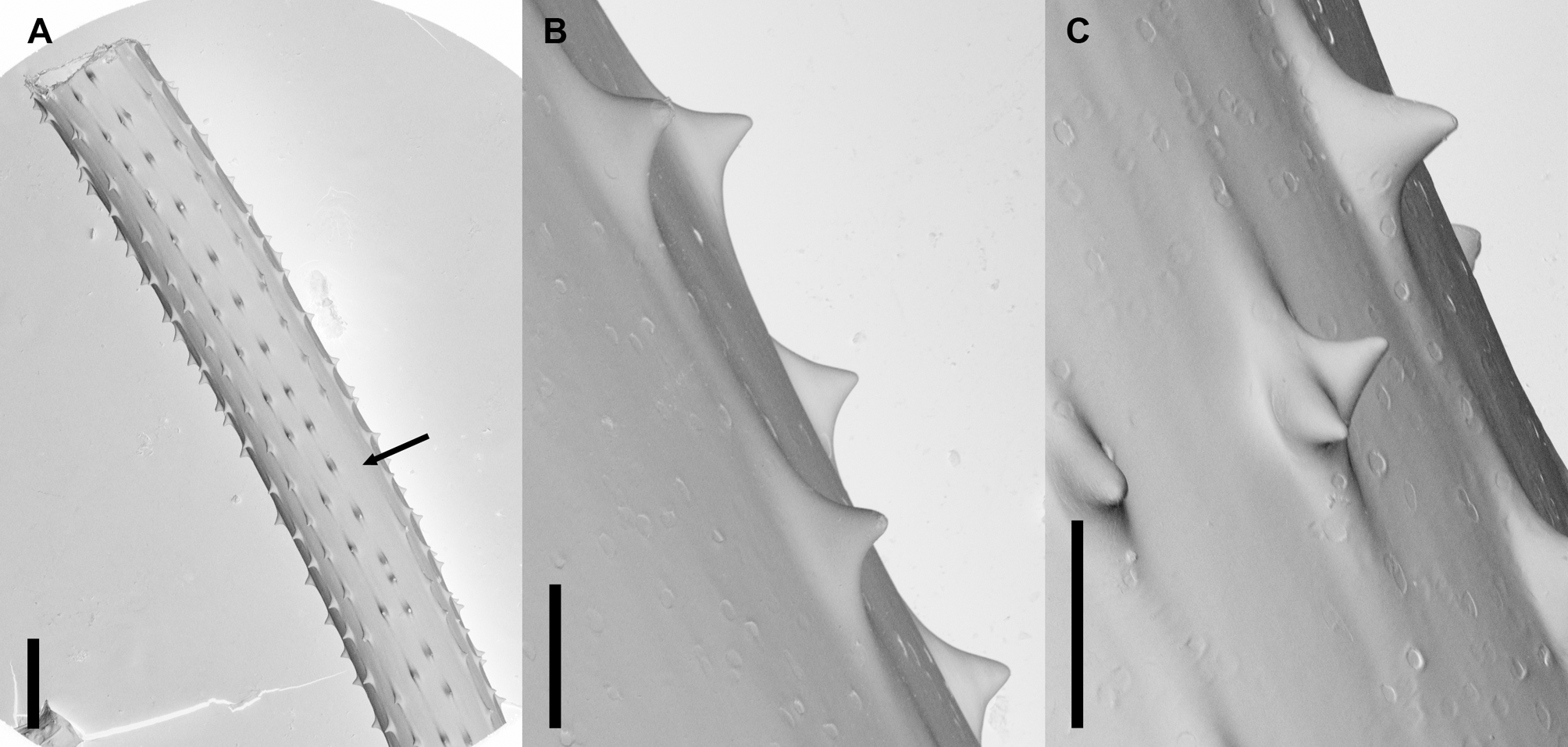
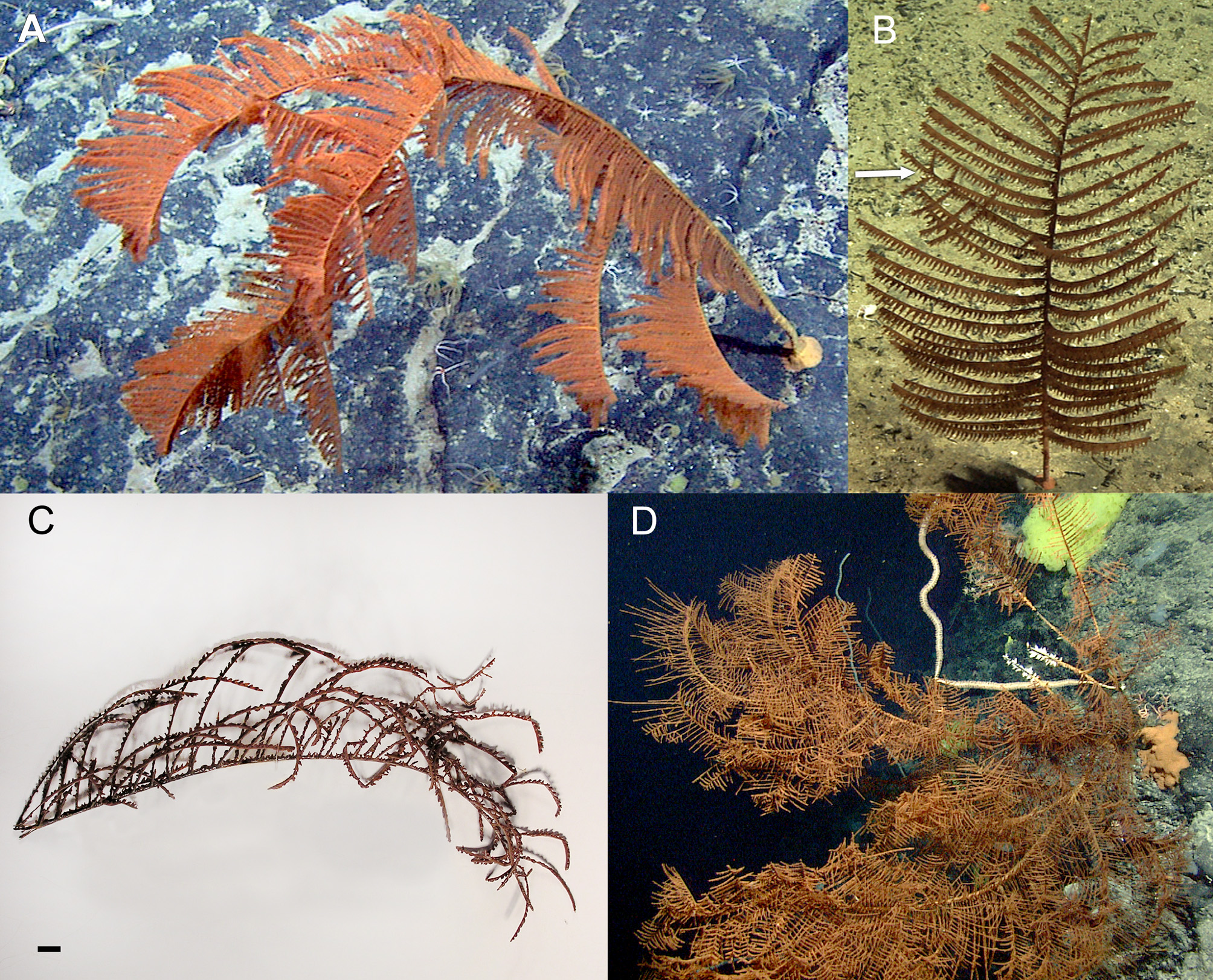
Description of the holotype. Telopathes magna spec. nov. was observed attached to a vertical cliff with a prominent stem and large, circular basal plate approaching 30 cm in diameter (Fig. 2 A – F). Orientation of the colony horizontal in the water, at a right angle to the vertical substrate. The type specimen is described from photos, video, and a fragment of one primary branch 16.7 cm in length, 36.0 cm wide. Fragment bearing two secondary branches 18.6 and 20.0 cm in length, 18.9 and 17.0 cm in width respectively (Fig. 3 A – B). Total colony size large, estimated at over 80 cm tall, greatest width approximately 85 cm. Stem mostly pinnulate, proximal few centimetres sparsely pinnulated or lacking pinnules. Stem thick near base, with an estimated basal stem diameter approaching 1 cm. Corallum branched to the second order, the longest branch approximately 39 cm long. Branches not restricted to one plane, directed distally. Pinnules long, 9 – 30 cm, shortening near branch tips, in two anterolateral rows, forming an acute angle between opposite pairs (Fig. 3 E). Pinnules directed distally. Branches more rigid while pinnules more flexible. One abnormally recurved pinnule observed in proximity to a deformed or bent secondary branch (Fig. 3 C – D), possibly indicating earlier damage to the colony. Living colony with bright orange polyps, tissue covering stem and branches more yellowish (Fig. 2 & 3). After preservation polyps brown, tissue lighter on the stems and branches. Tentacles long in living colony, up to an estimated 1 cm or more (Fig. 3 F), shorter after preservation. Polyps after preservation up to 7.1 mm in transverse diameter (Fig. 4 A – C). Skeletal spine morphology as described above, seven to nine longitudinal rows visible on pinnules in lateral view (Fig. 5 A – B), six to seven rows on branches. Rows on pinnules separated by 0.047 – 0.157 mm, spines within each row an average of 0.395 mm apart, not always uniformly spaced; occasionally sections in rows lack spines completely (indicated in Fig. 5 A), particularly in the distal sections. Occasionally spines are paired (Fig. 5 C) — a characteristic observed only on the pinnules, not on primary branches examined. Description of the Paratypes. Specimens from New England Seamounts similar morphologically to each other and holotype, but not identical, with branching to the first and second order in more than one plane (Fig. 6 A – D). All paratypes with relatively smaller basal plates compared to holotype, and basal plate not concentric on one large colony, indicating some intraspecific variation in this character, possibly due to the nature of the substrate on which the planula settles. Colony from Rehoboth Seamount (Fig. 6 D) appears to be the largest observed based on lasers on in-situ photographs, with conservative estimates of maximum height and width of approximately 133 cm and 150 cm respectively. The largest pinnules were estimated to be slightly smaller than the holotype at 24 cm in length. Colony branching was more dense than in smaller and probably younger colonies. Skeletal spines on pinnules collected from paratypes similar but not identical to holotype, are small and conical, with a size ranging from 0.019 mm (Rehoboth Seamount specimen) to 0.065 mm (Retriever Seamount specimen); smaller than those observed in the holotype. Distribution of spine sizes measured reveals a typically small difference between polypar and abpolypar sides. FIGURE 4. Polyps of Telopathes magna. (A – C) Arrangement of polyps along a pinnule; M = Mouth, ST = Sagittal tentacle, LT = Lateral tentacle. (A) Oral view. (B) Side view; P 1 & P 2 = individual polyps. (C) Close-up view of a typical polyp. (D) Arrangement of polyps along the primary branch. Scale 0.5 cm. The juvenile / small colony from Milne-Edwards Peak (Fig. 6 B – C) measured 27 cm in length, 13 cm in width, with the largest pinnule 7 cm in length. This specimen superficially resembles the genus Bathypathes. Primary pinnules large and simple, but are deflected anteriorly from the lateral plane. With one primary branch off main stem, this is distinguished by limited pinnulation and / or secondary branching. Skeletal spines on pinnules similar but not identical to holotype, small and conical, with a size ranging from 0.037 – 0.056 mm (Fig. 7 C). Distribution of spine sizes distinctly different on the juvenile colony, with notably larger spines on the polypar side, a range of 0.048 – 0.062 mm, and smaller spines on the abpolypar side, 0.037 – 0.047 mm with a mean of 0.048 mm (Fig. 7 C). All paratypes indistinguishable genetically from each other and the holotype. Comparisons. Telopathes appears morphologically most similar to the monopodial genus Bathypathes, especially smaller colonies, both possessing very long, simple pinnules in two rows, arranged in alternate or subopposite order. The polyps of both Telopathes and Bathypathes are large, the only two genera of Schizopathidae with polyps that may be in excess of 6 mm (Table 2). Differences include the branched corallum of Telopathes, though this branching may be slight, and the pinnules deflected from the lateral, observable even in the small colony (Fig 7 B – C). Schizopathes and Abyssopathes also possess large polyps and long, simple pinnules in two rows, but like Bathypathes, both are monopodial. In Schizopathes the pinnules are also lateral, not deflected, and the colony anchors in soft sediments, lacking a basal plate. Abyssopathes may have pinnules deflected from the lateral axis, like Telopathes, but also possesses the curious schizopathinid feature of one or more rows of anterior pinnules. Stauropathes is another large polyp genus, but the pinnules are short, and may be densely branched. The largest colony of Telopathes from Rehoboth Seamount was more densely branched than small and medium sized colonies observed, more similar to Stauropathes in this regard. It is possible that even larger / older colonies of Telopathes may be more densely branched, more closely resembling Stauropathes. All other genera of Schizopathidae belong to the subfamily Parantipathinae, with complex patterns of pinnulation (some combination of subpinnulation and / or additional rows of pinnules beyond two), and small polyps typically less than 3 mm in diameter.
Macisaac, K. G., Best, M., Brugler, M. R., Kenchington, E. L. R., Anstey, L. J., Jordan, T. (2013): Telopathes magna gen. nov., spec. nov. (Cnidaria: Anthozoa: Antipatharia: Schizopathidae) from deep waters off Atlantic Canada and the first molecular phylogeny of the deep-sea family Schizopathidae. Zootaxa 3700 (2): 237-258, DOI: 10.11646/zootaxa.3700.2.3