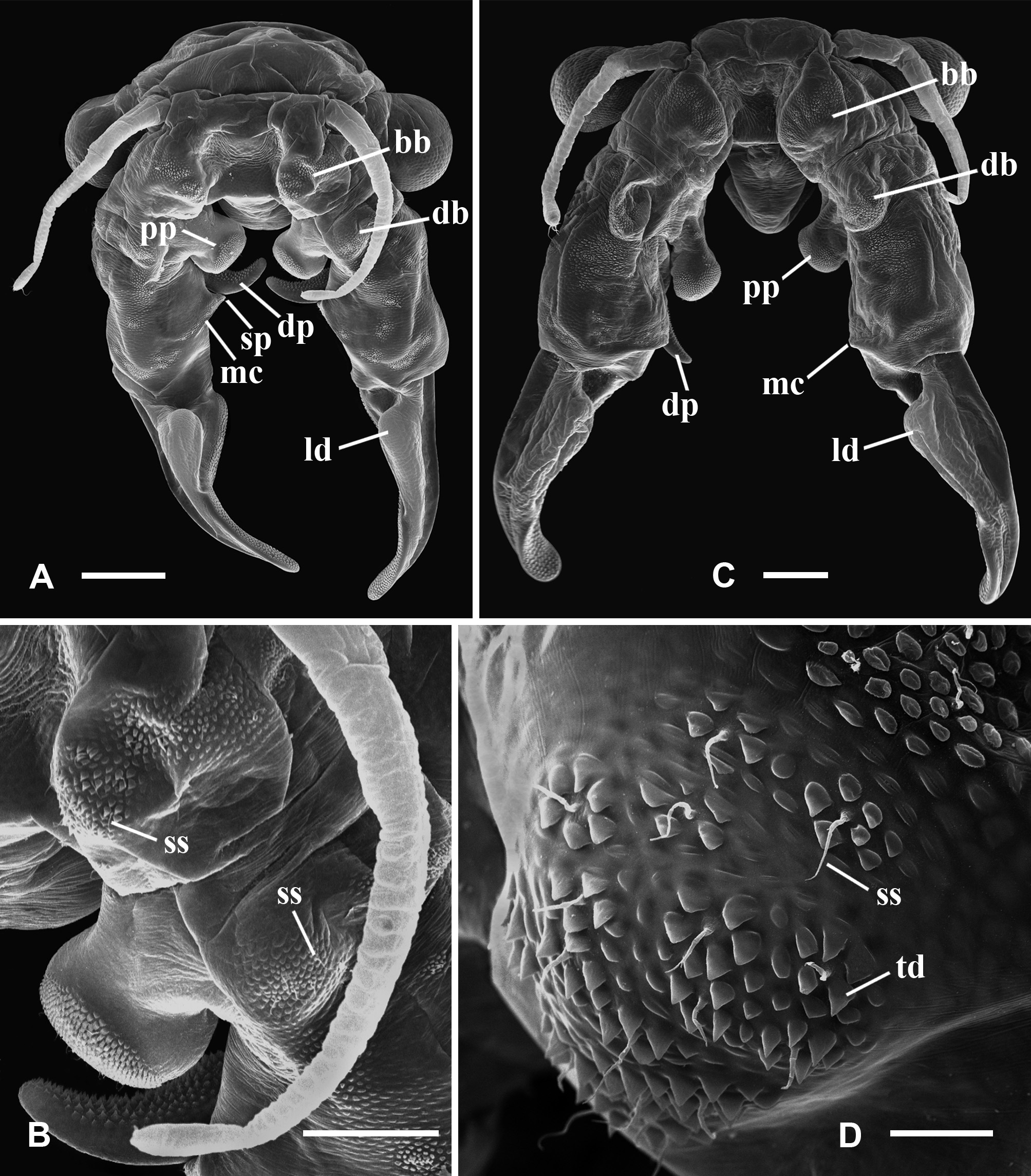
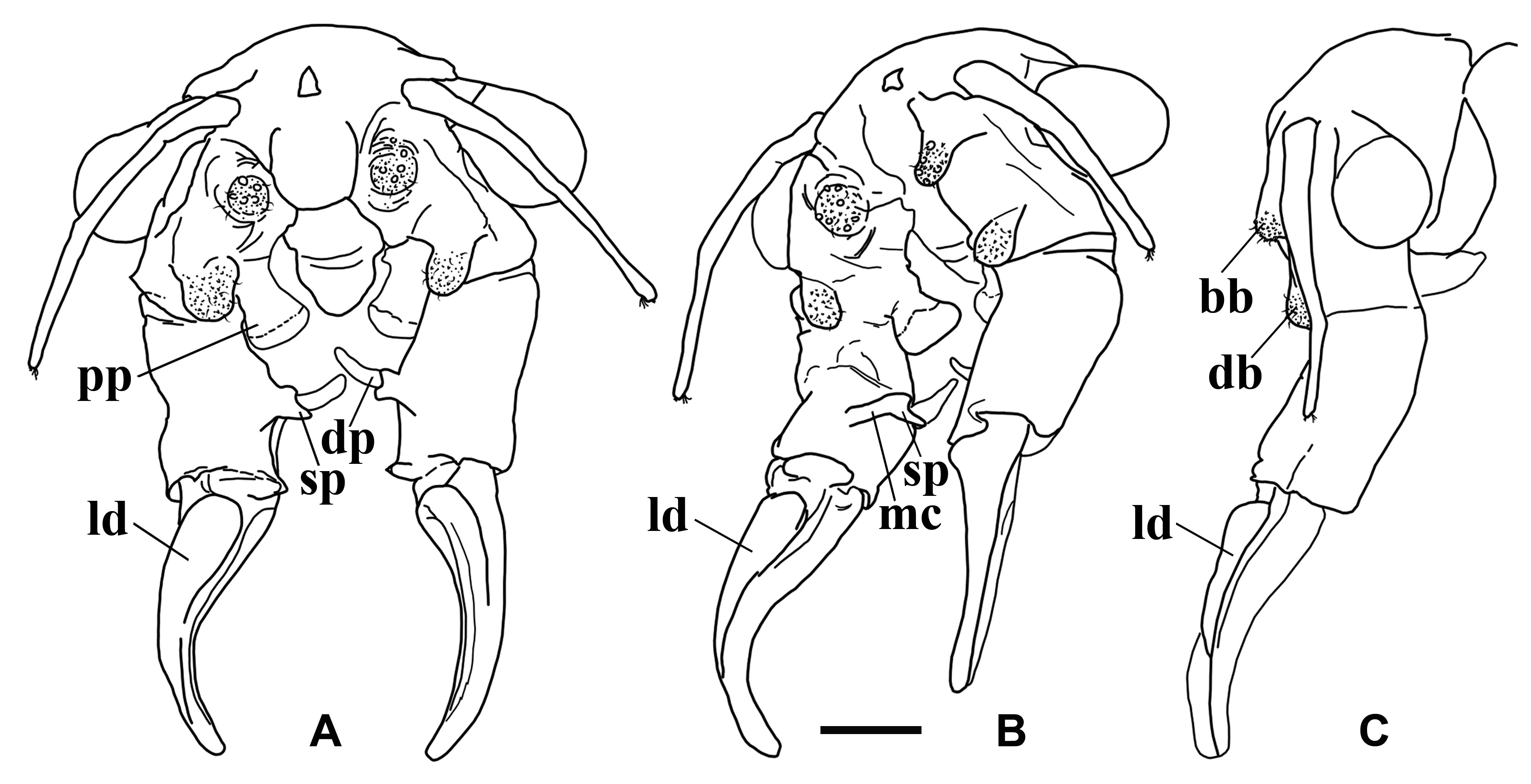
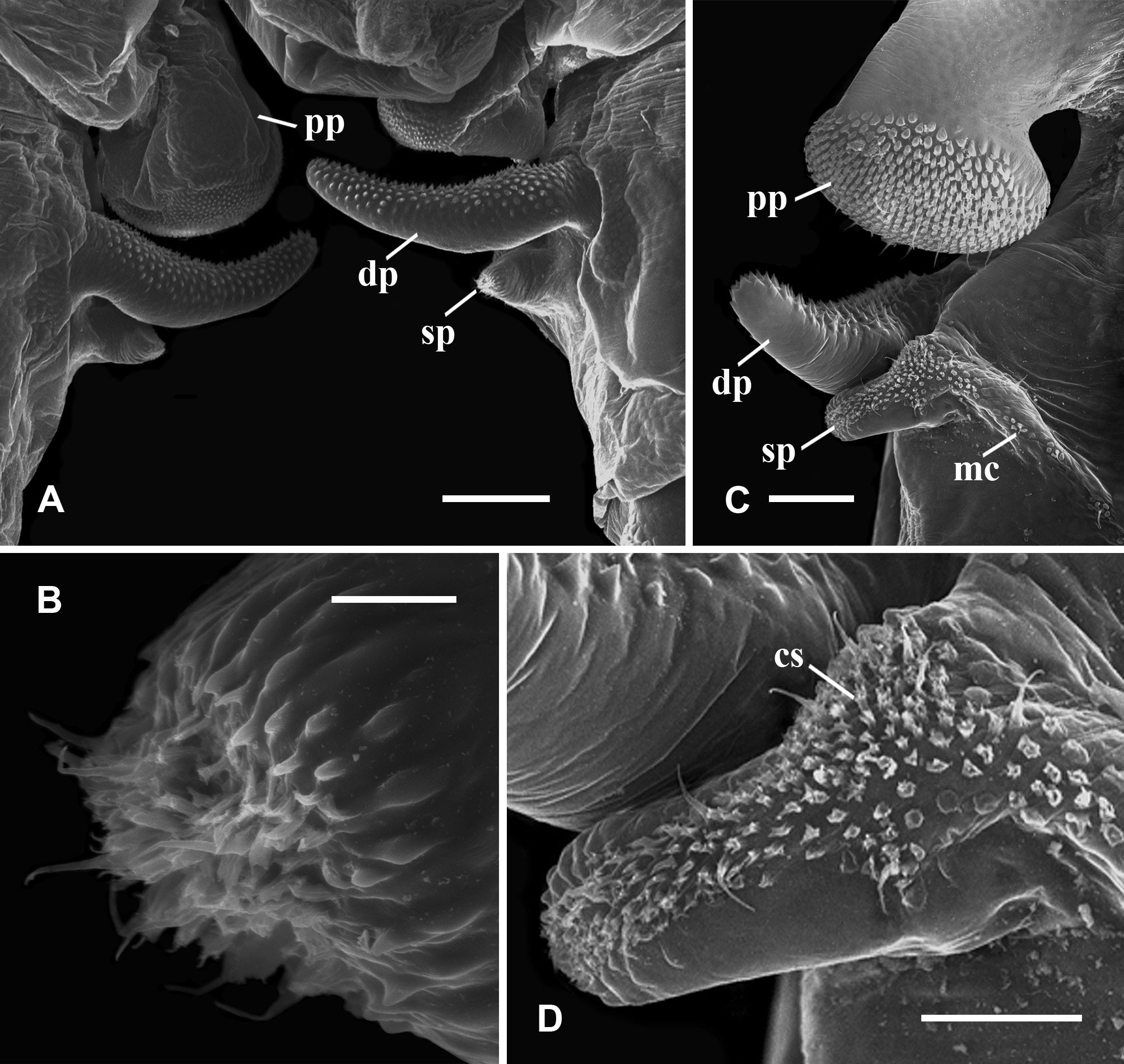
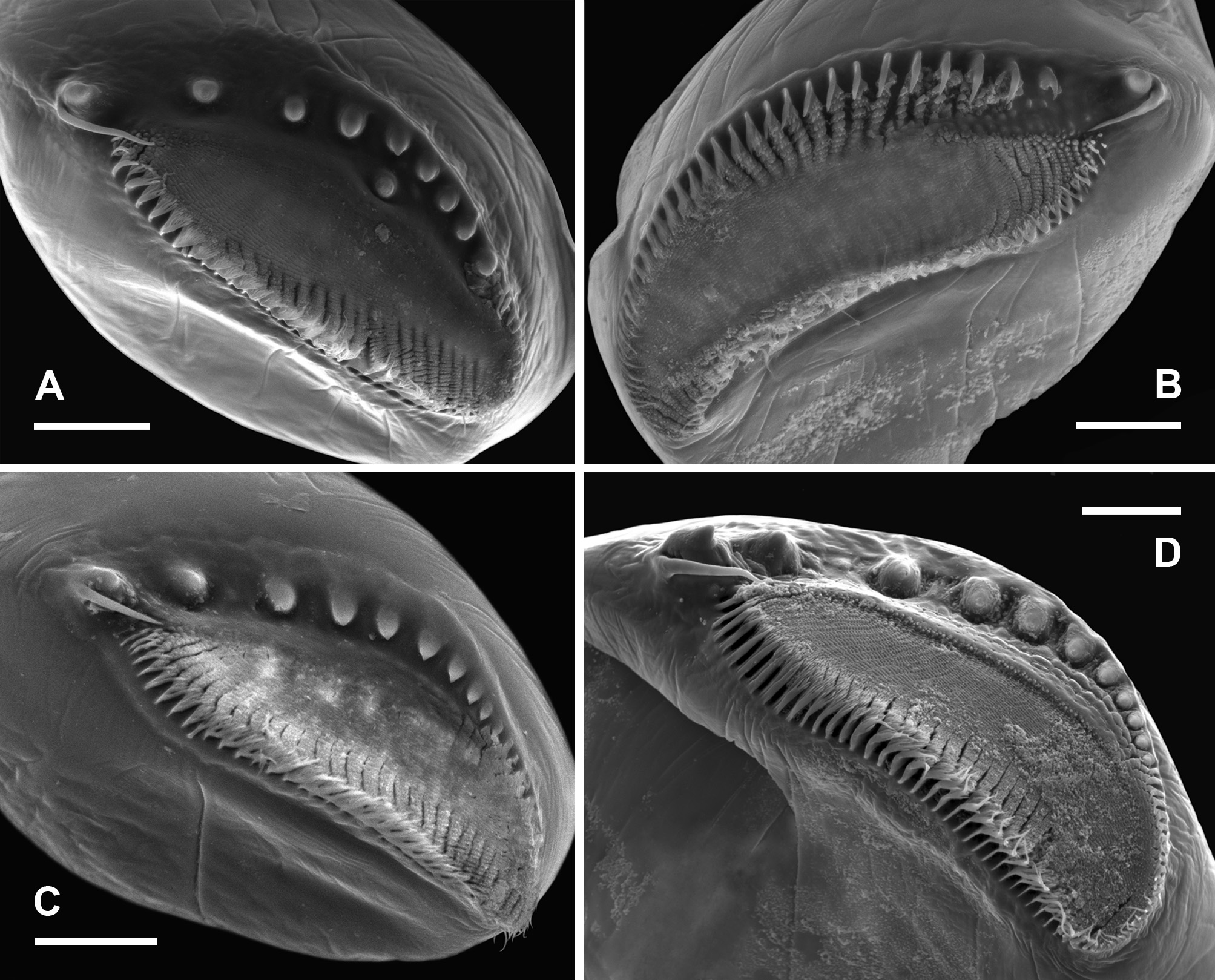
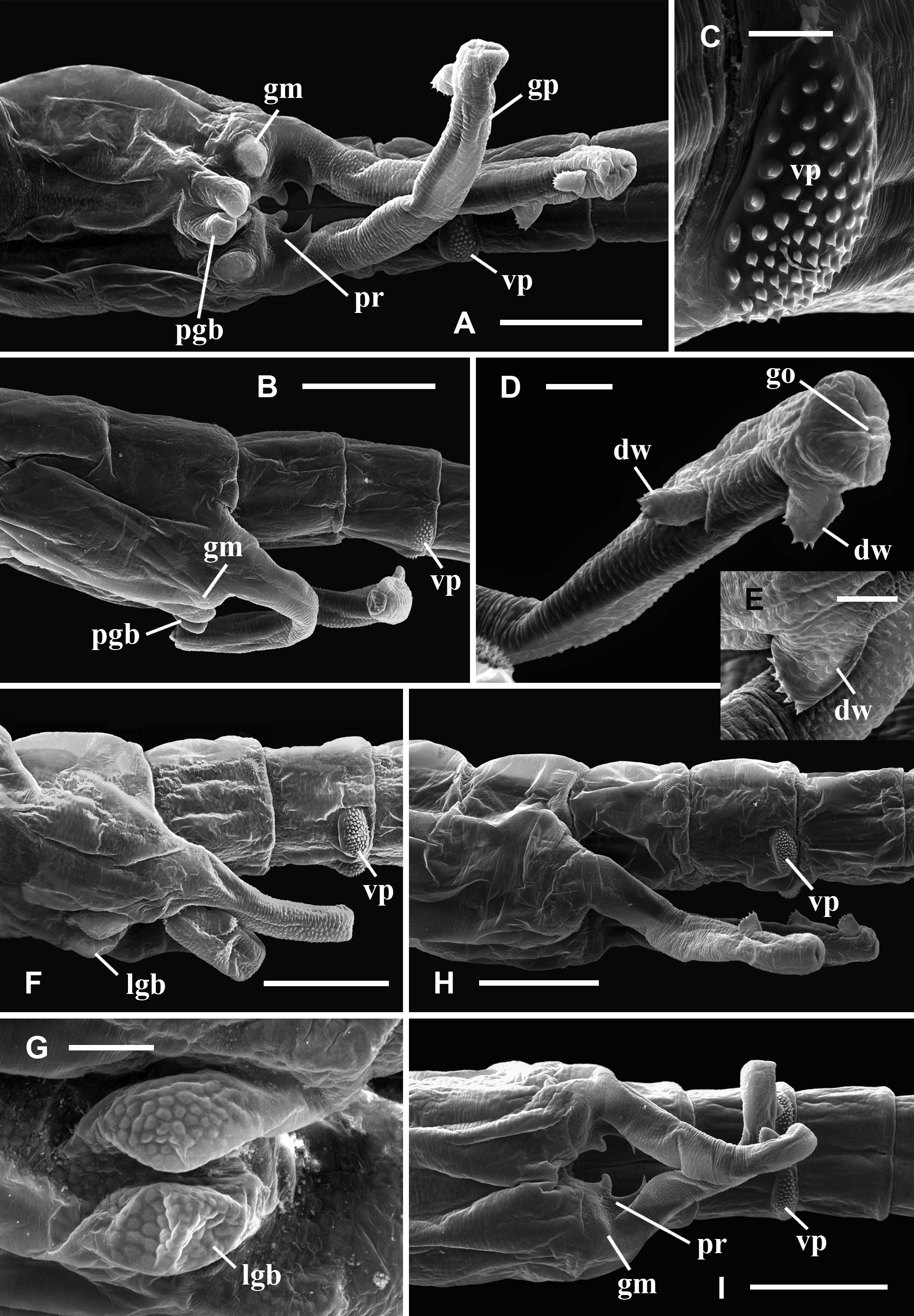
Redescription. Male. Cuticle ornamentation as in the present diagnosis of the genus (see below) .. Compound eyes mean diameter: 0.6 mm (0.4 – 0.6; n = 16), on average 1.3 times larger (1.2 – 1.5, n = 38) than in females (Tables 3, 4). First antennae partially multi-articulated with incomplete rings (Fig. 2 A, B). Second antennae basal antennomere, with a basal swollen bulge (bb), on the anterior surface, near the medial edge (Figs. 2 A, B; 3 A – C). The basal bulge is paved by small, slightly convex cuticular plates, some of them rise and point anteromedially, forming triangular denticles (td) arranged in circular groups around a central hyaline sensory seta (ss) (Fig. 2 B, D). The basal antennomere also bears on the anterior surface a more distal swollen bulge (db) (Figs. 2 A, B; 3 C), similarly paved with small convex plates which do not rise as denticles, and scattered warty sensory areas surrounding a hyaline seta (Fig. 2 B). Posteromedially at the same level, a spherical knob hangs from a peduncle as a pedunculate process (pp) (Figs. 2 A, B; 3 A; 4 A, C); the peduncle has a granulous cuticle, whereas the spherical tip is ornate with tightly arranged mesetiform elements with interspersed setae (Fig. 4 C). More distally in the basal antennomere, and also protruding from its posteromedial surface, there is a conspicuous digitiform process (dp), elongate, slightly curved, medially directed (Figs. 2 A; 3 A; 4 A, C), almost making contact in the midline with the digitiform process of the opposite side (Figs. 2 A; 4 A). The digitiform process is ornamented with sharp denticles that are more developed on the surface just opposite to the ornate tip of the pedunculate process; from this surface to both sides, denticles gradually flatten until disappearing, so that the cuticle appears almost smooth (Fig. 4 A, C). At the level of the origin of the digitiform process but on the medial surface of the basal antennomere there is a transverse oblique crest or medial crest (mc) (Figs. 3 B; 4 C) that protrudes posteromedially into a small process (sp) (Figs. 3 A, B; 4 A, C, D), usually ending in two rounded posteromedial tips, one more prominent than the other, adopting together a cordiform shape; the small process seems to prevent the digitiform process of its own side from moving beyond; the small process is ornamented with columnar grooved spines (cs) distally divided into several irregular points, scattered simple smooth spines, and interspersed hyaline sensory setae (Fig. 4 B, C, D). The second antenna distal antennomere bends from its anterior surface in posteromedial direction, ending in a rounded spatulate tip. A little beyond its proximal articulation, the anterolateral surface of the antennomere protrudes like a vertical globose triangular ledge (ld) with proximal base (Figs. 2 A; 3 A – C), which narrows in distal direction and ends up at the distal third of the lateral edge of the antennomere. A wide rasp-like area covered with conspicuous mesetiform ornamentations, is observed from the tip of the appendage to the posterior and medial surfaces of the distal antennomere. Mandibles asymmetric, showing no sexual dimorphism (Fig. 6 A, B); maxillae 1 and 2 as typical in Branchinectidae. Thoracopods as in the present diagnosis of the genus (see below). Genital and abdominal segments. Abdominal segment II (second postgenital segment) as herein redescribed in the genus, bearing a pair of ovoid ventrolateral denticulate protuberances (vp), close to its posterior articulation, showing diverse degrees of separation from each other in different specimens (Fig. 7 A, B). Pairs of similarly ornate but less prominent areas were observed in equal positions, near the posterior articulation of abdominal segments IV and VI. Besides, both genital segments present a conspicuous pair of dorsal warty sensory areas, also observed in several abdominal segments. Gonopods (gp) widely separated, extending ventrolaterally, with their non-eversible portions partially visible in dorsal view; very long when completely everted, reaching abdominal segments III or the beginning of IV (Fig. 7 A). The rigid basal portion of gonopods bears a medial sclerotized projection with two processes (pr) (Fig. 7 A); in addition, laterally in the basal portion there is a ventral pair of granulous mounds (gm) (Fig. 7 A, B). In ventral view, near the distal edge of the second genital segment and between the rigid basal portions of both gonopods, there is a pair of protruding genital bulges (pgb), distally rounded, and next to each other at the midline; in lateral view they are conspicuously prominent (Fig. 7 A, B). Each eversible portion of the gonopods shows two dentate warts in its distal part (Fig. 7 A). Both warts are forwardly and obliquely directed, and bear 6 to 8 short marginal spines; one of the warts is medial and close to the tip of the gonopod and the other is dorsal and located at three quarters of the length of the everted portion of the gonopod (Fig. 7 A). No anterior or lateral longitudinal rows of denticles were observed on the retractile part of the gonopod. Internal structure of the male genital segments (Fig. 8 A) as described in the present diagnosis of the genus (see below). Testes reach abdominal segments I to III (Fig. 8 A). Uropods (or cercopods) length: 1.4 mm (1.0 – 1.7 mm; n = 16), slightly longer than in females (male uropod / female uropod: 1.2 (1.0 – 1.4); n = 38), on average 3 times as long as the anal segment (or telson) (2.6 – 3.8; n = 16) (Tables 3, 4), divergent, uniformly provided with marginal plumose setae. Female. Cuticule ornamentation as in male. Compound eyes smaller than in male, mean diameter: 0.4 mm (0.4 – 0.5 mm; n = 25) (Table 4). First antennae as in male. Second antennae as in the genus, presenting a cheliform aspect (Fig. 9 B); indeed, the specific epithet “ pollicifera ” proposed by Harding (1940) alludes to this character. The anterior surface of the antenna as well as the medial projection (mp), including its tip, present warty sensory areas (sa) associated with a central seta (Fig. 9 F). Instead, the apical projection (ap) of the appendage lacks sensory areas. In some locations the specimens show a pair of medial bulges (mb) (Fig. 9 B). Mandibles as in male (Fig. 6 A). Maxillae 1 and 2 as typical in Branchinectidae. Thoracopods as in the present diagnosis of the genus (see below). Thoracic ornamentation. Thoracomers 9 or 10 to 12 (first genital segment) showing a pair of dorsal evaginations confluent at the midline, close to the posterior articulation of each segment, which gradually increase in size in the posterior direction (Figs. 10 A, B). In segment 9, when present, confluent evaginations end in two close rounded tips; in thoracomers 10 to 12, the confluent evaginations end in two close conical points; sometimes, the strongest conical dorsal points in the first genital segment show laterally a conspicuous sensory area with an associated seta or with a little spine. Thoracomer 13 (second genital segment) with a ventrolateral strong spine (sg 2), curved, posteriorly directed, just above the brood pouch (Fig. 10 A, B, C), and a pair of dorsal warty sensory areas associated with a central hyaline seta, which may be either a conspicuous dorsal mound or barely prominent. Abdominal segment II bearing a pair of ovoid ventrolateral denticulate protuberances (vp) (Fig. 10 E) as in males (Fig. 10 A), which may be hidden by the brood pouch; similar but smaller structures were also observed in abdominal segments IV and VI. Ovaries T-shaped, extending to abdominal segments I – III, or rarely to IV – V. Brood pouch subcylindrical, rounded at the tip with the gonopore (go), bearing at its distal third a pair of small ventrolateral cuticular rounded outgrowths (lo) with cuticular granulations as in the whole brood pouch (Figs. 10 A, C, D). Brood pouch reaching abdominal segments II – IV, or more rarely the middle of V. Uropods (or cercopods) length: 1.2 mm (0.8 – 1.8 mm; n = 25), slightly shorter than in male (Tables 3, 4), on average 2.4 times as long as the anal segment (or telson) (1.7 – 3.0; n = 25), divergent, uniformly provided with plumose marginal setae (Table 4). Cysts (resistant eggs): two kinds of cysts were found in the brood pouches of Archaebranchinecta, being type I the most common in both species. Type I cyst in A. pollicifera: spherical, mean diameter: 246.4 µm (196 – 262 μm; n = 11), surface sculptured with ridges defining numerous cells (9 to 11) across the egg diameter, cells irregularly sized, subcircular or oval, mean dimensions: 23 – 30 µm (18 – 34 to 29 – 32 µm; n = 24), either isolated or coalescent (more frequently in pairs up to 5 cells delimited by narrower and lower ridges), cell groups either lined up or form- ing diverse arrays (since type I cysts are similar in both species, Fig. 11 A corresponds to A. aimara); each cell forms a shallow depression with grained bottom (Fig. 11 C); ridges steep-sided and narrow except at intersections. Type II cyst: found together with type I cyst in Pe- 29 and Pe- 30, at the northern reach of our sampling area. Spherical (Fig. 11 B), mean diameter: 204 μm (174 – 248 μm; n = 6), surface sculptured with ridges defining few large cells (6 to 8) across the egg diameter, cells irregularly sized, subcircular or oval, mean dimensions: 37 – 47 µm (24 – 38 to 61 – 67 µm; n = 22), either isolated or coalescent; with flat-bottomed grained depressions (Fig. 11 D); wide ridges with flat tops and sloping sides. Size. male total length from the front to the tip of uropods (or cercopods) excluding setae: 12 mm (8 – 14 mm; n = 16); measurements of other characters in Table 3. Female total length from the front to the tip of uropods (or cercopods) excluding setae: 13 mm (10 – 19 mm; n = 25); measurements of other characters in Table 4.
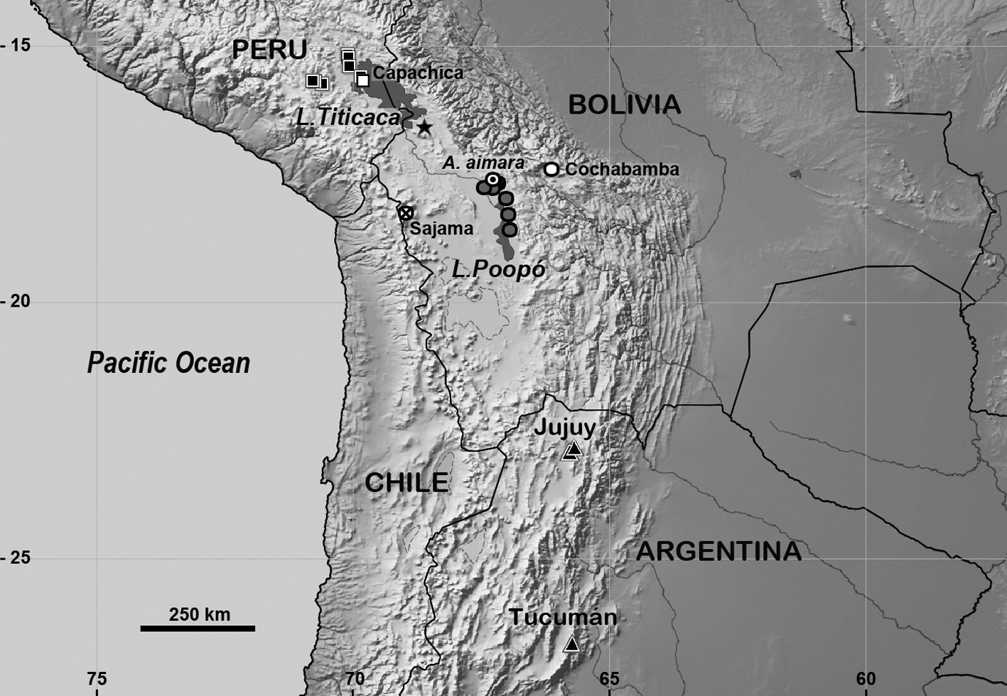
Cohen, Rosa Graciela, Marinone, María Cristina, Adamowicz, Sarah J. (2019): A new species of Archaebranchinecta (Anostraca: Branchinectidae) from the South American Altiplano. Zootaxa 4683 (1): 55-83, DOI: 10.11646/zootaxa.4683.1.3