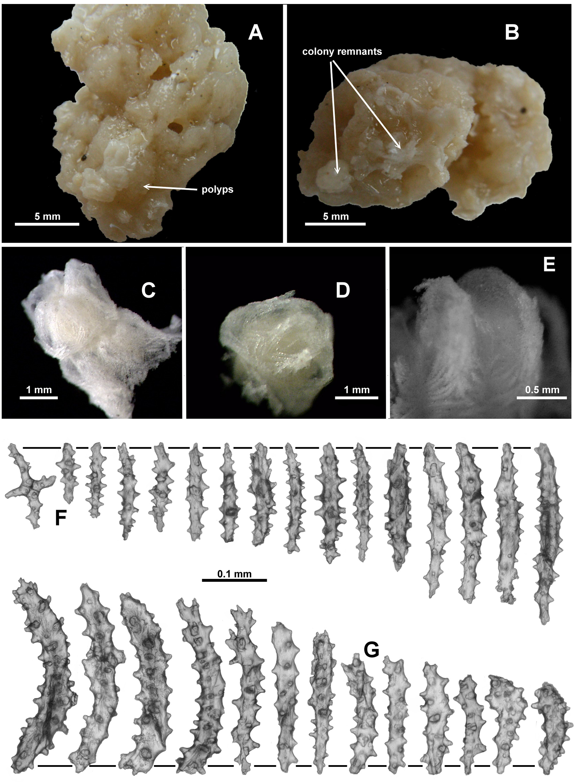
Description: Colony form: Unfortunately, there is nothing remaining of the syntype lodged at the Museum of Natural History, Wroclaw University, Poland (MZW # 49), only the sponge on which it grew. No further mention is made of this syntype. The lectotype is a tiny, encrusting colony growing on a small cream sponge (Fig. 16 A, B). The piece of sponge is approximately 20 mm long and 15 mm wide with the octocoral colony growing on only a small part of it. The colony is difficult to discern on the very similar coloured sponge but there are very few remaining polyps and they are quite damaged. Three polyps are grouped close together at one end of the sponge. They appear to be attached to one another via stolons and a spreading membrane as mentioned by Kükenthal (1913), with no discernible branching. Colour: Kükenthal (1913) states the colour as light yellow. Polyps and calyces: Calyces are prominent and approximately 2 mm tall (Kükenthal mentioned 2.5 mm tall) formed from a thin layer of sclerites (Fig. 16 C). Some appear to have rounded longitudinal furrows but they are not pronounced. In most of the remaining polyps, the polyp body is partly retracted such that the base of the polyp head rests on the lip of the calyx, hiding the polyp neck. The polyp heads are approximately 1.5 – 1.7 mm wide and polyps are approximately 1.5 – 2 mm tall (2.5 mm tall in Kükenthal’s description). There are some smaller polyps, possibly juveniles (0.9 mm wide and 1.2 mm tall). The tentacles fold over the mouths of the polyps forming eight distinct furrows on the polyp head and a star-liked apex to each polyp. The number of pinnules per tentacle could not be determined. Medulla and Cortex: No medulla evident. Sclerites: All polyps, calyces and colony surface are covered in a dense layer of sclerites, chiefly sticks and spindles. Sclerites on the polyp head are in a clearly defined collaret and points arrangement (Fig. 16 D). At the collaret, sclerites are basically transverse but grade en chevron up the points to become longitudinal in the distal part. These sclerites are short, straight or only slightly curved rodlets and spindles with fairly sparse, conical tubercles (Fig. 16 F). The lengths of the sclerites are 0.1 – 0.3 mm (mostly 0.1 – 0.2 mm) only, making them significantly smaller than those found in other Anthothela species. Distal from the points, sclerites continue longitudinally to obliquely, along the aboral side of the tentacles (Fig. 16 E). They are curved rodlets and spindles, some slightly clavate, with sparse, conical tubercles (Fig. 16 G), and they grade in size from largest at the proximal end of the tentacle to smallest distally and seem to curve around the sides of the tentacles. Tentacle sclerites are 0.14 – 0.34 mm long which again is smaller than the range found in other Anthothela species but the ranges do overlap. In the pinnules are found very short, almost squat, spatulate clubs, with a narrow handle and a flattened, wide tip, along with numerous short rods (Fig. 17 A). The spatulate tip is oriented distally in the pinnules and some of them have quite well developed ‘ teeth’ or jagged edges. Spatulate clubs range from 0.09 – 0.16 mm only — much smaller that the equivalent spatulate clubs in A. grandiflora where the range is 0.26 – 0.5 mm. The rodlets are short and narrow, with simple, small tubercles, and are also crowded longitudinally in the pinnules (Fig. 17 A). They range in size from 0.06 – 0.18 mm. On the calyx, sclerites are arranged at all angles in a relatively thin but crowded layer. Sclerites again are straight, short rods and spindles with sparse tubercles, ranging in size from 0.16 – 0.34 mm (Fig. 17 B). One straight club with a thickened distal tip was found (Fig. 17 Ba), which corresponds to a sclerite figured by Kükenthal (1913), although he states it is from the top part of the polyp. No samples of a pharynx were examined, so the presence, absence or nature of sclerites in the pharynx remains undocumented. Surface sclerites are a mixture of two sorts: short, straight, tuberculate rods and spindles (0.07 – 0.18 mm long) (Fig. 17 C) and longer, straight sticks and spindles, mostly smooth or with minimal tubercles, sometimes with forked ends (0.13 – 0.24 mm long) (Fig. 17 D). The latter are reminiscent of the sclerites found in the medulla of other species of Anthothela. Variability: A small fragment of the specimen USNM 57981 was examined. The largest portion of the colony consists of four narrow branches emanating from a membranous base with some anastomoses evident; it has no main stem and calyces are distributed throughout but also tend to form terminal bunches (Fig. 17 E). There is an additional fragment (Fig. 17 F) that has well-defined ridges on the calyces and most of the polyps partly retracted so only the top part of the polyp head is visible in the mouth of the calyx. The fragment examined was a small branch tip with a few polyps crowded together (Fig. 18 A). The tallest polyp is partly extended and is 1.5 mm tall and 1.6 mm wide (Fig. 18 B) while the calyces are approximately 1.5 – 2.5 mm tall and 1.5 – 1.8 mm wide. Under a dissecting microscope the sclerites are opaque and white and they are brown under transmitted light, perhaps indicating the specimen was originally fixed in formalin. There is slight damage evident in some of the sclerites. The fragment available for examination is small, and the only medulla portion available is affected by the junction of a polyp, so it is impossible to be definite regarding coelenteric canals penetrating the medulla and even the expected boundary canals are distorted and difficult to discern. Nevertheless it is clear there is an axial medulla and a cortex. The pinnules have numerous spatulate clubs, many having a long, narrow handle with a wide, flattened spatulate tip, and they are longer than those of the lectotype (0.12 – 0.22 mm); but there are also some short, squat spatulate clubs similar to the lectotype (Fig. 18 C). The small, narrow rodlets from the pinnules are of similar length in this specimen (0.04 – 0.18 mm) as in the lectotype. The tentacles have mostly curved, short, narrow tuberculate rods, arranged longitudinally on the aboral side of the rachis (Fig. 18 D). These are very similar to the lectotype in form and length (0.14 – 0.35 mm). Sclerites in the points are long tuberculate sticks and spindles, slightly curved or straight (Fig. 18 E), plus some straight sclerites with a clubbed, thorny tip (Fig. 18 Ea) which are not present in the small sample from the lectotype. Most point sclerites are significantly larger than those from the lectotype (0.19 – 0.68 mm cf. 0.1 – 0.28 mm). This specimen had a low number of sclerites in the pharynx, mostly short, simple spindles varying in length from 0.07 – 0.19 mm (Fig. 19 A). Most of the sclerites from the calyx are tuberculate sticks and spindles (Fig. 19 B) although there are some clavate sclerites which resemble the clubbed sclerite from the lectotype (Fig. 17 Ba). These are all a similar size to the lectotype at 0.14 – 0.38 mm long. The cortex sclerites are similar to those from the calyx although with fewer clavate sclerites (Fig. 19 C). Most of the cortex sclerites are 0.12 – 0.24 mm long but there are also longer sclerites, up to 0.44 mm long. Occasionally, straight clubs are found in both the cortex and the calyx. As mentioned above, there is very little true medulla available in the fragment investigated and no sclerites similar to those found in the medulla of other Anthothela species were observed.
A taxonomic revision of Anthothela (Octocorallia: Scleraxonia: Anthothelidae) and related genera, with the addition of new taxa, using morphological and molecular data