
Corella eumyota
Chileense zakpijp(+16)·Traustedt, 1882
GBIF:5200463
0
Descendants
0
Synonyms
0
Children
PROFILE
Species Profile
ABOUT
Descriptions(9)
Corella eumyota Traustedt, 1882 from Chile
Figs 3, 4A.
Material examined
- 42°27’ S – 73°45’ W, 24/08/1972, coll. Coloma.
- Chili without precision, 26/03/1970, coll. Gallardo. - Guaitecas Islands, 44° S–073° W, 24 m, SMNH 126199, Arnback-Christie-Linde coll.
The type of Corella eumyota from Chile (Valparaiso) could not be found among the Traustedt material (pers. com. Zoologisk Museum Copenhagen).
We have examined Corella samples present in the MNHN collections collected close to the type station and they correpond well to the Traustedt description. Ärnback-Christie-Linde (1929) described from Chili (Guaitecas Islands) small specimens with the same musculature and gonads as in C. eumyota but the gonoducts are not mentioned. We examined her specimens and they probably belong to the same species as the ones described by Traustedt. They are from a near by area. In her discussion she already doubted of the identity with true Antarctic specimens. The anatomical characters correspond to the Traustedt description.
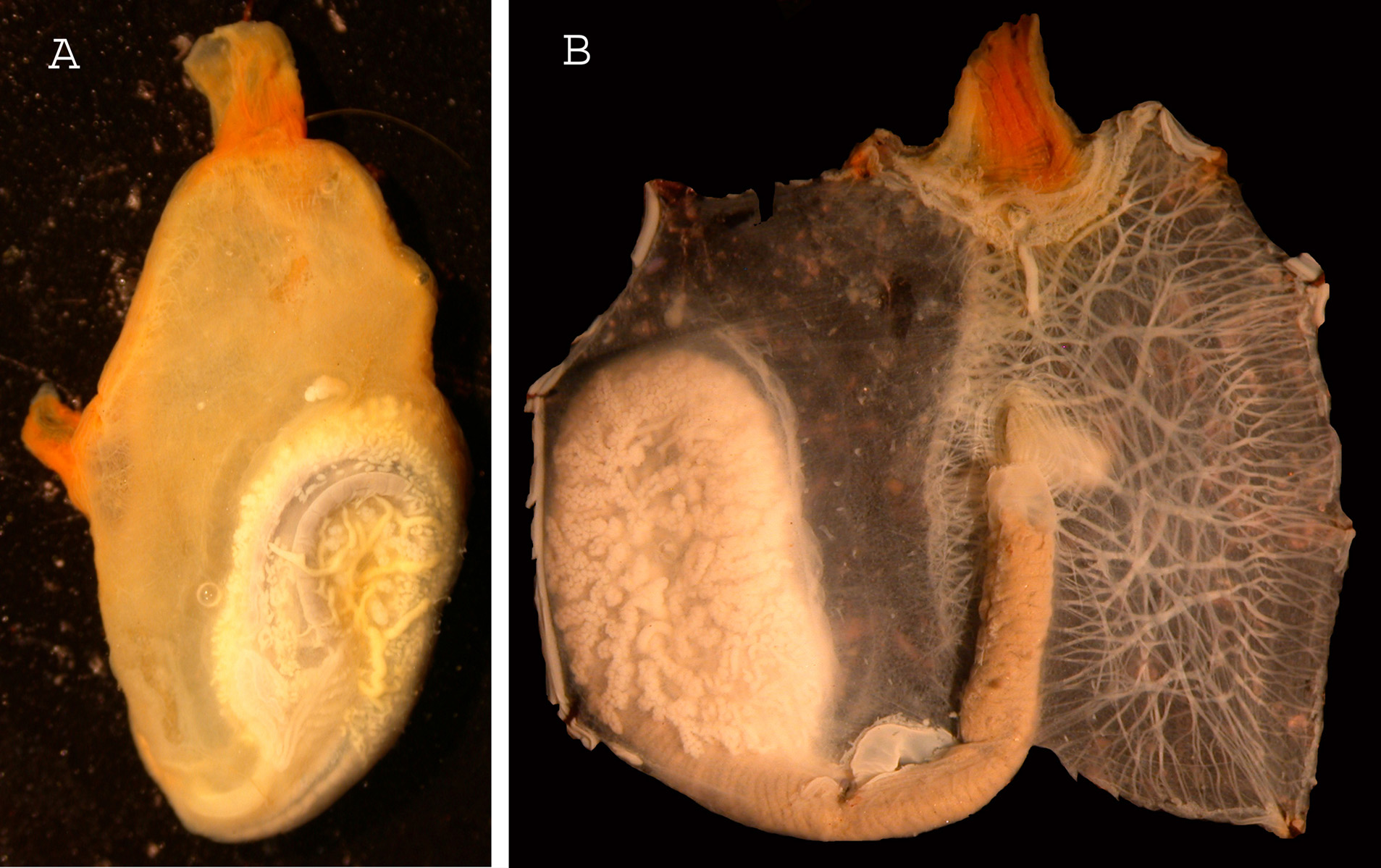
Some of the specimens mentioned above are aggregated; the largest is 4.5 cm long. The tunic is translucent when fixed. It has a rather smooth surface but wears some thin papillae and a few epibionts. Its consistency is cartilaginous. The oral siphon has 6 low lobes and is terminal and protruding with ocellli. The atrial siphon forms a short tube at 1/3 of the body length; its rim has also 6 lobes and ocelli. The animals were fixed by the right side. The tentacles are numerous in 3 orders of size. Their number varies according to the size and the stations. The dorsal tubercle is U-shaped and anteriorly open. The pre-pharyngeal band dorsally curves to become a groove anterior to the dorsal languets. The musculature on the siphons is strong. The longitudinal fibres coming from the oral siphon extend along 1/5 to 1/4 of the body length on both sides. On the left side transverse muscular bundles start from the ventral line and divide to make a dense network with the transverse ribbons issued from the dorsal side (Fig. 3 A). The body wall around the gut has no muscles (Fig. 3 A).
The branchial sac is flat, and thin (Fig. 3 B). The dorsal lamina is attached to the body wall and to the intestine ending at the level of the oesophagus entrance. The dorsal languets are equal in length less numerous than the transverse vessels, and inserted on a flat imperforated lamina strongly linked to the rectum. There is an average of 35 longitudinal sinuses on each side, thin and rarely interrupted. The stigmata are not regularly lined. The spirals (Fig. 3 B) are irregular in 2-3 interrupted turns and often subdivided, the number of turns is lower in the part of the branchial sac located over the gut.
The digestive tract is large and occupies a large part of the right body side (Fig. 3 A). The oesophagus entrance has a thick lip. The olive-shaped stomach has longitudinal folds hidden internally by the gonads. On the external side of the cardia, the typhlosolis can be more easily seen. The intestine draws a vertical closed loop and curves below the oesophagus to join the dorsal line to which it is attached along its whole length. The anus has a circular outline with small round lobes (Fig. 3 D). The testis vesicles are irregularly but mostly grouped on the external side of the intestinal loop and included among pyloric vesicles. The male ducts sink inside the gut and are hardly visible, opening on the internal side of the gonad by a simple fringed hole (Fig. 3 C arrow). The oviduct opens close to the male aperture in a wide hole with small round papillae on its rim (Fig. 3 C arrow).
Eggs were present into the atrial cavity, but no tadpoles have been found.
Corella eumyota from Sub-Antarcic regions and Europe
Figs 5, 6
Material examined
- New Zealand: Portobello: shore and wharfs, coll. A.Wood, 2012. Dunedin, coll. Lambert, 16/01/1994. - Amsterdam Island, 37°50 S–77°30 E, 80m, 11/01/1972, coll. Beurois. - South Africa, Hout Bay, 7/02/1996, coll. Monniot.
- France: Le Havre harbour, 2010, 2011, 2012, coll. Breton; Fecamp 2007, coll. Breton; Camaret 2002, coll. Lambert; Arradon 2009, coll. Le Roux.
Turon (1988) partially described and represented specimens identified as C. eumyota from Namibia (lagoon, 1m depth) but the specimens are lost (Turon pers. com.).Their tunic is said to present “small tubercles” and the stigmata have only 1 or 2 spiral turns. The gonads (Turon 1988 Fig. 5) are limited to the bottom of the gut loop, The genital ducts are not figured nor mentioned. This species may be C. eumyota .
Brewin (1946) described as C. eumyota ascidians from New Zealand and precised that some of them were slightly different with a long sperm duct. She probably overlooked the presence of a second species C. brewinae n.sp. in addition to C eumyota .
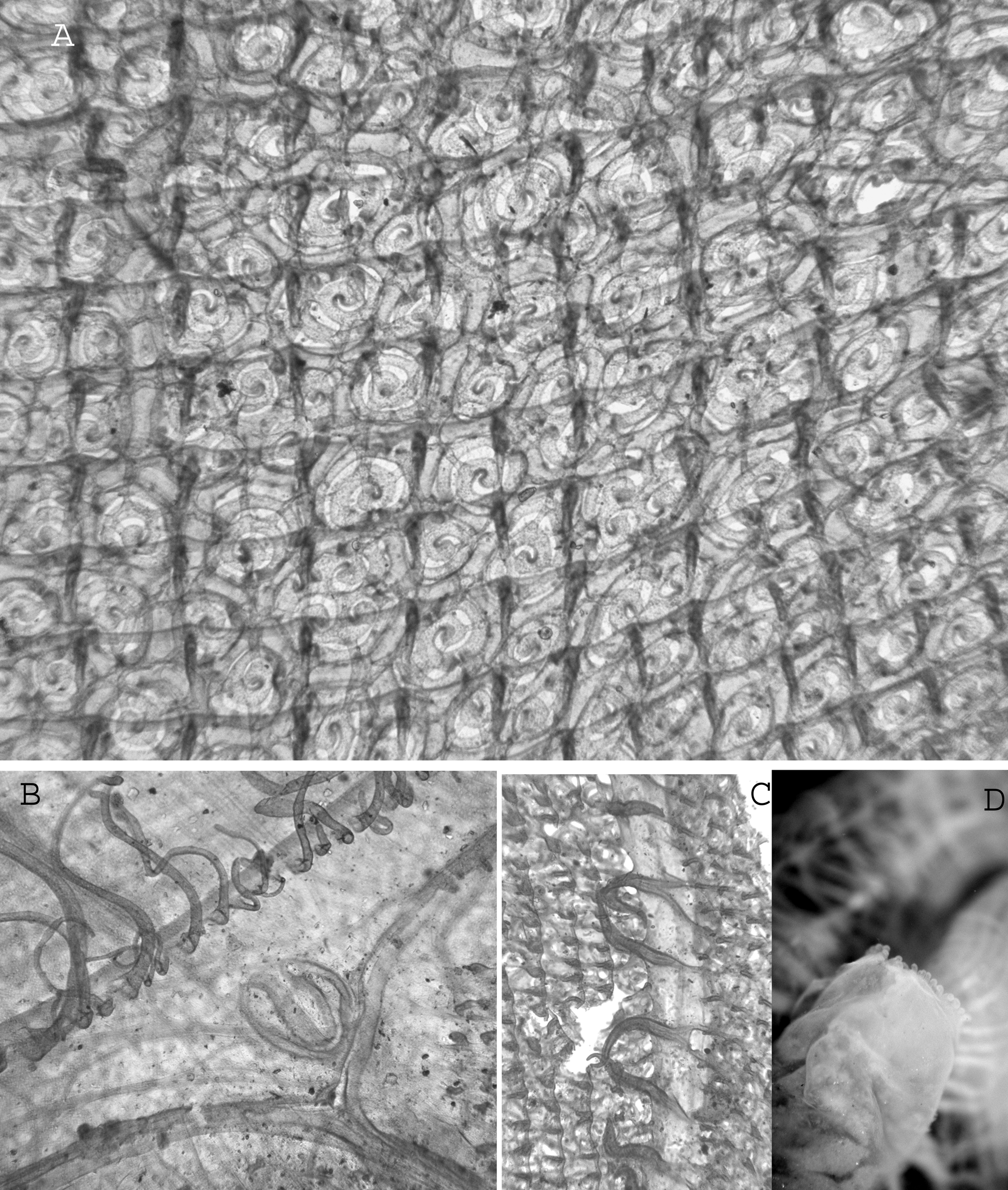
There is a dense population of Corella developed in the littoral area in the vicinity of Portobello (New Zealand) and it has been known for a long time. Specimens newly collected from Portobello and specimens collected by Lambert (1994) from Dunedin have been compared with the Chilean ascidians. The results are absolutely similar: body proportions (Fig. 5 A), muscular design (Fig 5 B), dorsal tubercle (Fig 6 B), branchial tissue (Fig. 6 A) dorsal languets (Fig. 6 C), anus (Fig. 6 D), genital papillae. Similarly no differences could be found with the numerous specimens collected along the French coast. (coll. Breton).
Export occurrence data
Darwin Core Archive (ZIP)
GEOGRAPHY
Distribution Map
REGIONS
Geographic Distribution(96)
DATA
Occurrence Datasets
Common names used for this species across different languages and regions. Available in 4 languages and 2 countries. 1 preferred.
Vernacular (common) names are the everyday names used for a species in different languages and regions. A single species may have dozens of common names worldwide. This taxon has names in 4 languages. 1 name preferred.
CLASSIFICATION
Taxonomic Classification Tree

HIERARCHY
Child Taxa(2)
RELATED
Related Name Usages(20)
Matching names from other GBIF-indexed checklists and datasets.
MULTIMEDIA
Media Files(5)
FIGURE 7. A, Ciona robusta on a buoy, note also some Asterocarpa humilis (orange-coloured). Corella eumyota. B, whole individual; C, dissected individual; D, right side of the mantle, branchial sac eliminated. Inset shows magnification of the zone of the genital openings. The specimen in C and D has been stained. Scale bars: A, 10 cm, B, 2 mm, C, D, 1 mm.

FIGURE 3. Corella eumyota specimen from Chile. A, body ventrally opened; B, branchial tissue; C, genital papillae (arrows) on the internal side of the gut loop; D, anus.
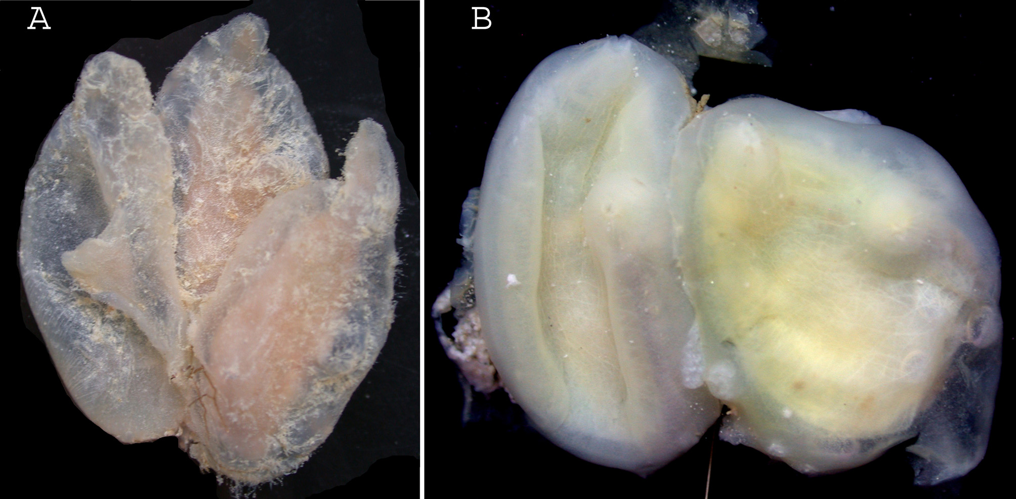
FIGURE 4. A, Corella eumyota aggregated specimens from New Zealand; B, Corella brewinae n. sp. aggregated specimens from New Zealand.
IMAGES
Gallery(5)
Occurrences with images
CITATIONS
References(16)
Alurralde, G.; Torre, L.; Schwindt, E.; Castilla, J. C.; Tatián, M. (2013). A re-evaluation of morphological characters of the invasive ascidian Corella eumyota reveals two different species at the tip of South America and in the South Shetland Islands, Antarctica. <em>Polar Biology.</em> 36(7): 957-968.
Alurralde, G.; Torre, L.; Schwindt, E.; Castilla, J. C.; Tatián, M. (2013). A re-evaluation of morphological characters of the invasive ascidian Corella eumyota reveals two different species at the tip of South America and in the South Shetland Islands, Antarctica. <em>Polar Biology.</em> 36(7): 957-968.
Clarke, A.; Johnston, N.M. (2003). Antarctic marine benthic diversity. <em>Oceanography and Marine Biology: an Annual Review.</em> 41: 47-114.
Clarke, A.; Johnston, N.M. (2003). Antarctic marine benthic diversity. <em>Oceanography and Marine Biology: an Annual Review.</em> 41: 47-114.
Hansson (1998) NEAT (North East Atlantic taxa): South Scandinavian marine Chordata Check-List.