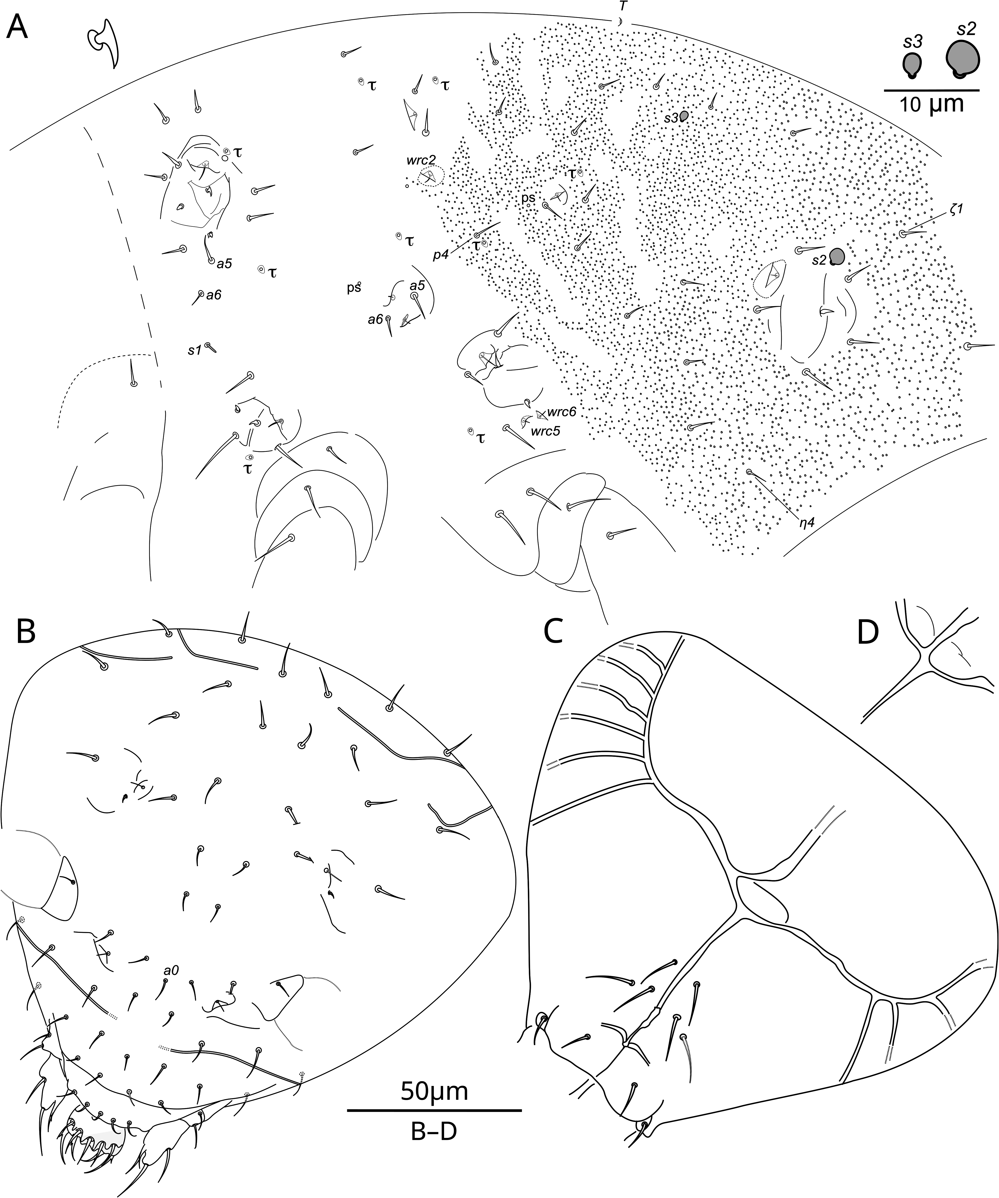
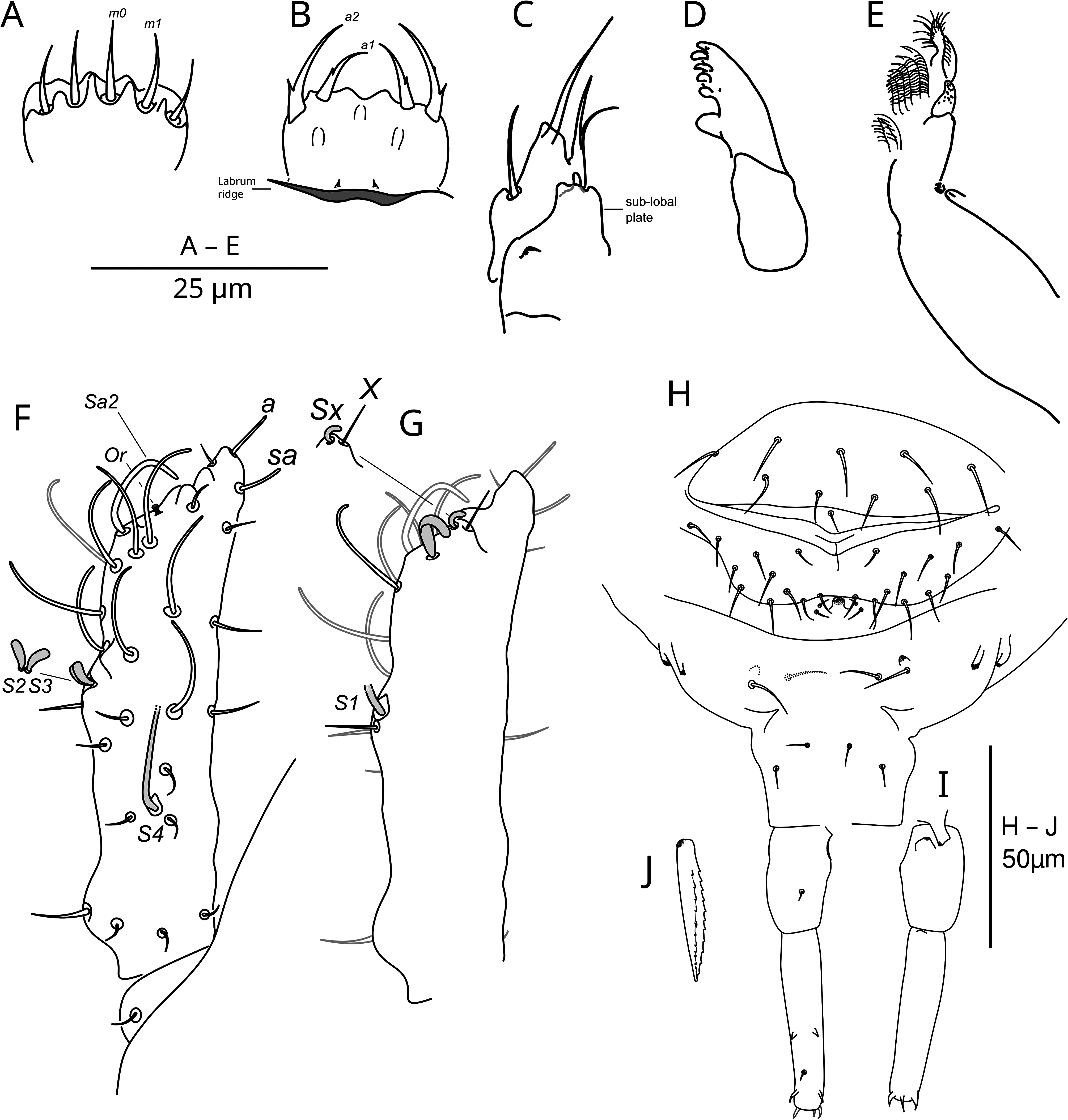
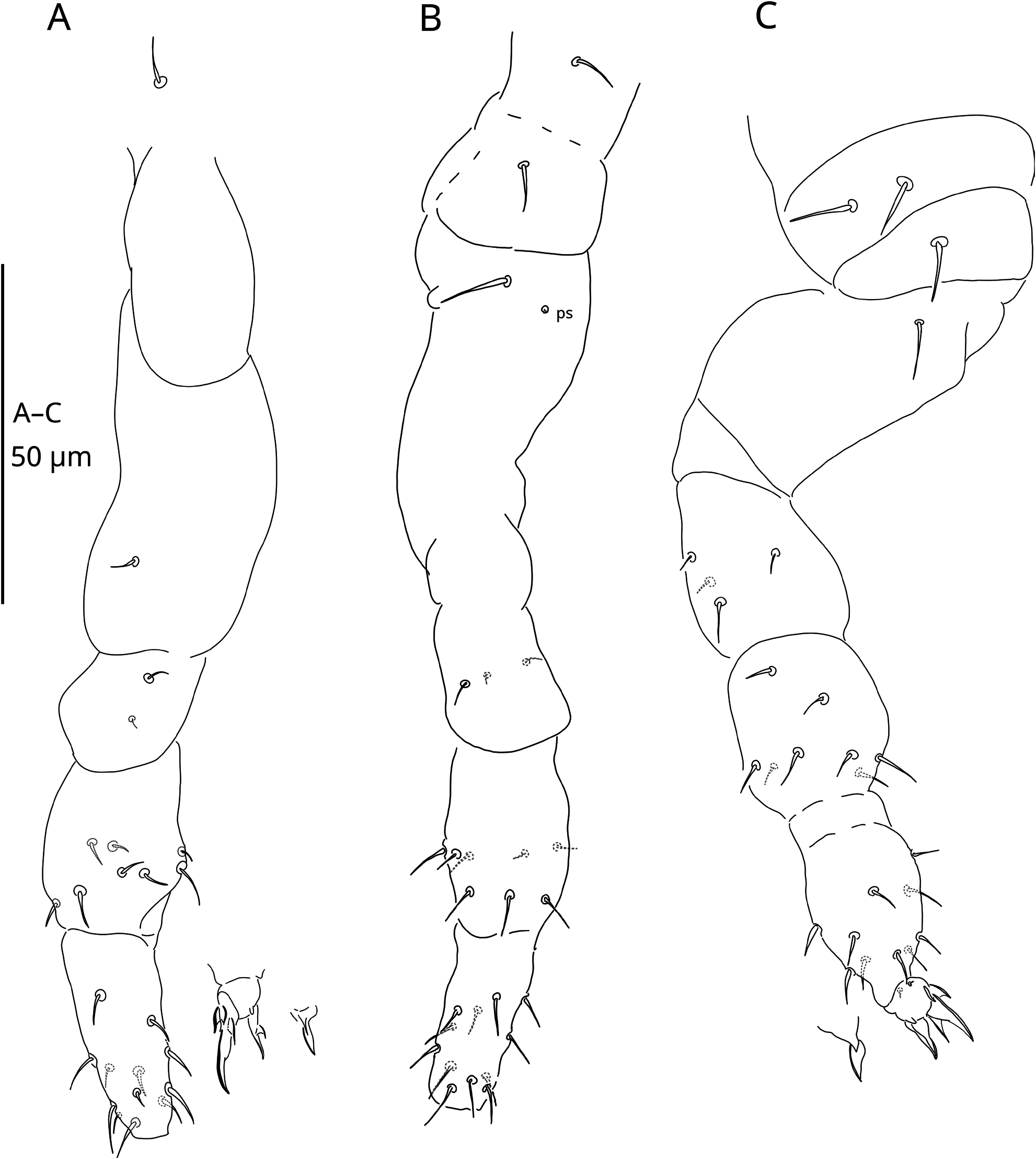
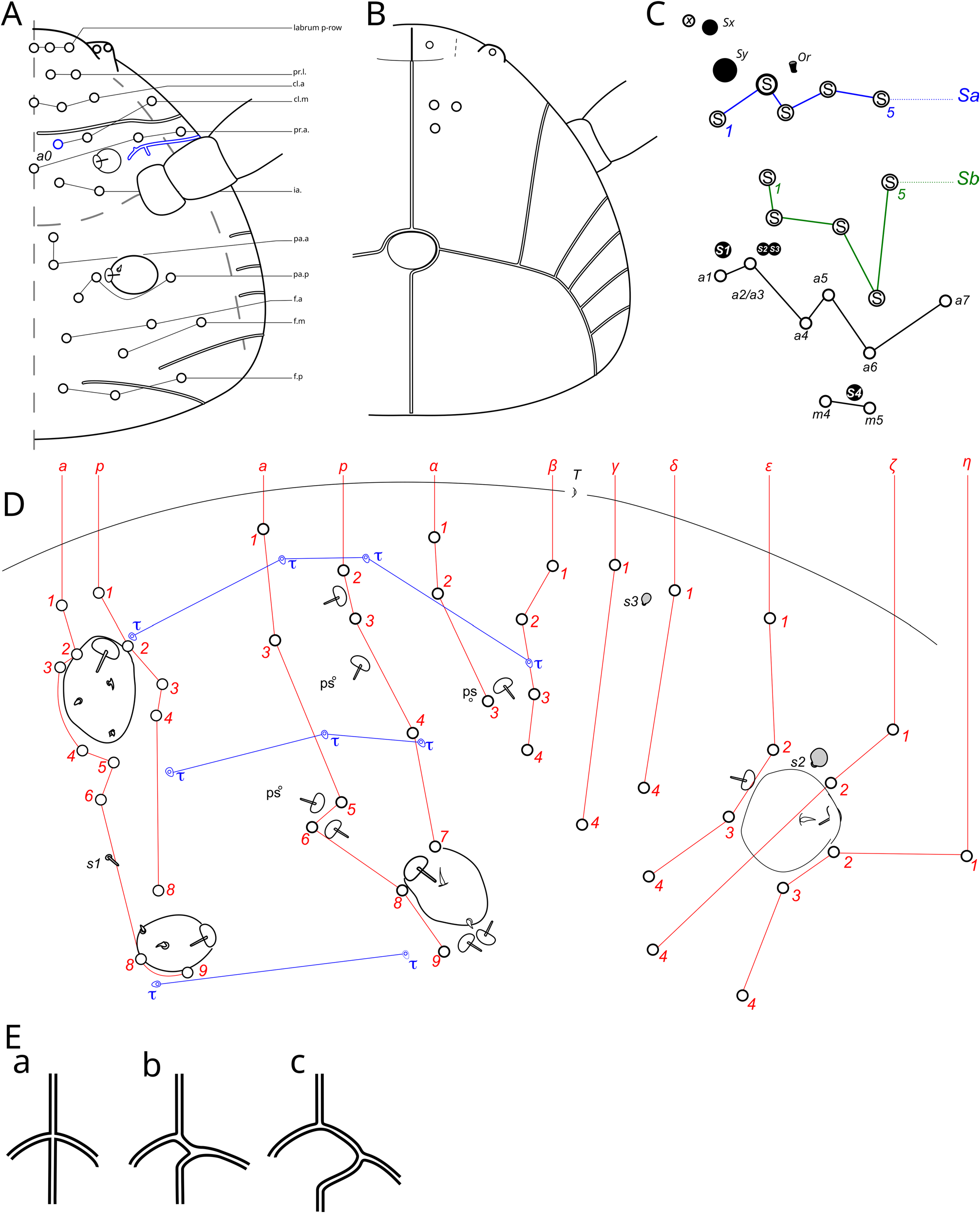
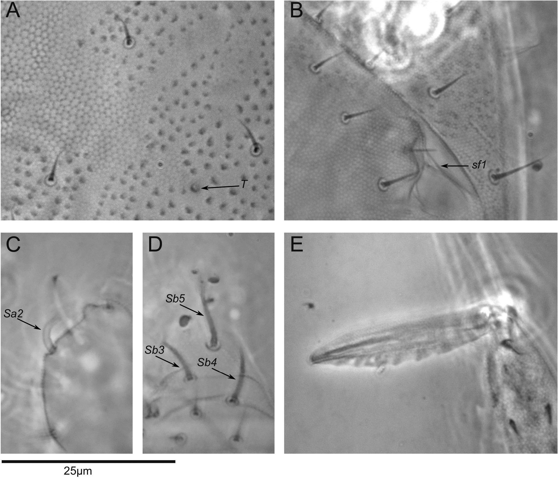
Description. General aspect. Habitus and segmentation typical of the genus. Length from labrum to anus: up to 450 µm. Specimens whitish to red (in 96 % ethanol). All typical chaetal types of the genus are accounted for, without any remarkable development. Integument. The ordinary secondary granulation is limited to the posterior half of the trunk (roughly corresponding to the abdominal region), absent on the head, the thoracic region and Abd. VI (Fig. 1 A). Anteriorly on the abdomen, dorso-median line also without secondary granulation until the terminal secondary granule T (same as in Fig. 9 A). Primary granulation pattern distinctly visible dorsally on the head and the thorax: primary grains stronger and hexagons surface larger than on the secondary granulated part of the abdomen (same as in Fig. 9 A). On the clypeal area, a speckled pattern of hexagons with enlarged primary grain yielding superficial resemblance to ordinary secondary granulation (same as in Fig. 9 B). Integumentary channels moderately developed on the head (Fig. 1 B, C). On each side of the head, the basal channel splits successively. At each split, the external branch is always terminal (never splitting). The first external branch extending to the antero-dorsal region of the head, then up to four branches extending to the latero-posterior part of the head. Finally, the two most posterior branches reaching the dorso-posterior part of the head. Channels connection with linea ventralis from circular (mostly observed, Fig. 1 C) to almost crossed (seen in one specimen, Fig. 1 D). Channels absent on trunk. Sensory fields and wax rods. Ordinary distribution of sensory fields and wax rods secretory crypts: 2 + 2 wrc on head, 12 + 12 wrc on body; including the ones associated with the 6 + 6 sensory fields (Fig. 1 A, B). Sensory fields include the swollen inner chaetae in the usual distribution (from sf 1 – 6: 0, 1, 3, 2, 2, 1). The swollen inner chaetae are all short and flame-shaped, some with a tendency toward the T-shape (Fig. 1 A). Level of separation of wrc 5 and 6 from sf 5 cannot be quantified with the amount of secondary granules (non applicable). Head chaetotaxy. Postero-dorsal chaetae without remarkable thickening, only subtly stronger than antero-dorsal chaetae (Fig. 1 B). Number of chaetae: 12 + 12 in the postero-dorsal region, 8 + 8 and 2 unpaired in the antero-dorsal region, 2 + 2 in the antero-lateral region (Fig. 1 B, 8 A). Ventrally, 3 + 3 chaetae in the sub-labial region (Fig. 1 C, 8 B). Diagram of head chaetotaxy provided in Fig. 8 A, B. Labium. Basomedian fields of labium with 1 + 1 chaetae, basolateral fields of labium with 1 + 1 chaetae on a small papilla (Fig. 1 C). Labial palps ordinary. Labrum. Chaetae a 1 and a 2 thicker than chaetae m 0 - 2; a 1 shorter than a 2; m 0 - 2 smooth; a 1 and a 2 acuminate, with inward curvature, each with one external, basal tooth (Fig. 2 A, B). A median, very fine tooth can sometimes be perceived on a 2 (Fig. 2 B). Labral anterior process apparently as in M. minimus, with a continued transversal crest separating a 1 - 2 from m 0 - 2. Ridge of the labrum with at least two small teeth (Fig. 2 B). Other mouthparts. Oral fold with 2 + 2 chaetae (Fig. 1 B). Maxilla outer lobe with two chaetae (apical and basal) and with a strong bifurcate hair in subapical internal position (Figs 1 B, 2 C). Sub-lobal plate without hair, but with a small lobe near the anterior ridge (Fig. 2 C). Mandibula with the ordinary asymmetry: one strong basal tooth on the left mandibula (Fig. 2 D), missing on the other side. Maxilla with a well developed apical lamella (Fig. 2 E). Antenna. Ant. I with one chaeta (Fig. 2 F). Ant. II with four chaetae, the anterior one stronger than the three others (Fig. 2 F). Ant. III with eight chaetae and five S-chaetae including S 1 – S 4 from the sensory organ and Sb 4; S 2 and S 3 small but clearly protruding from the cupule; without perceptible ornamentation in light microscopy (Fig. 2 F, G). Ant. IV with five chaetae (X present) and 11 S-chaetae (Sb 1 – 3, Sb 5, Sa 1 – 5, Sx and Sy); Sa 2 is notably enlarged in regard to the others common S-chaetae Sa and Sb (as in Fig. 8 C); apical and subapical rods (a, sa) present (Fig. 2 F, G). Diagram of antennal chaetotaxy provided in Fig. 8 C. Thoracic tergites. Th. II with 12 + 12 ordinary chaetae, 1 + 1 s-chaetae s 1 and three τ-chaetae (only the bases of τ-chaetae could be perceived using light microscopy); chaetae a 5, a 6 present and chaeta a 7 missing; the laterodorsal τ-chaeta is far from chaeta p 4, in the lateral direction (Fig. 1 A). Th. III with 10 + 10 ordinary chaetae, 6 + 6 wrc, 4 + 4 τ-chaetae and 2 + 2 pseudopores (ps); a 5 sensibly bigger than a 6, wrc 2 distant from p 4 (Fig. 1 A). Diagram of chaetotaxy provided in Fig. 8 D. Abd. I – V tergites. With 22 + 22 ordinary chaetae, 2 + 2 globular s-chaetae (s 2, s 3) s 3 notably smaller than s 2, 2 + 2 τ-chaetae, 1 + 1 pseudopores and 2 + 2 wrc; chaetae ζ 1 and η 4 present (Fig. 1 A). Abd. VI. Tergite with 4 + 4 and 1 unpaired chaetae. Each anal valves with one small chaeta. Sternite with 10 + 10 chaetae (Fig. 2 H). Genital plate. Ordinary: female with 2 + 2 chaetae (Fig. 2 H). Males present, but the genital plate could not be studied in detail. Abd. IV sternites. With 2 + 2 usual neosminthuroid chaetae, usually with 2 + 2 ordinary chaetae and 1 + 1 small lobes but anomalous asymmetry observed in one specimen (Fig. 2 H). Abdominal appendages. Manubrium with 2 + 2 posterior chaetae (Fig. 2 H). Dens ordinary: basal part of dens with 1 + 1 posterior chaetae, apical part of dens with 1 + 1 posterior chaetae and 7 + 7 small spines (Fig. 2 H, I). Mucro elliptical, with moderately wide lamellae, with a dozen of teeth on each posterior lamellae (Fig. 2 J). Ventral tube with 2 + 2 apical chaetae, retinaculum with 3 + 3 teeth. Legs. Chaetal composition on each legs subcoxa 1, 2, coxa, trochanter, femur and tibiotarsus: Leg I, 1, 0, 1, 2, 8, 12 chaeta (e) (Fig. 3 A); leg II, 1, 1, 1, 3, 8, 12 chaeta (e) (Fig. 3 B); leg III, 2, 1, 1, 4, 8, 10 chaeta (e) (Fig. 3 C). Claws ordinary, as in Fig. 3 A, C., with short external basal lamellae.
A new group of species of the genus Megalothorax (Collembola, Neelidae) with Gondwanan distribution, and introducing an open interactive identification key of Megalothorax species