
Anyuta anastema
Grischenko, Gordon & Melnik, 2018
GBIF:10339847
PROFILE
Species Profile
Habitat
Characteristics
ABOUT
Descriptions(5)
Export occurrence data
Darwin Core Archive (ZIP)
CLASSIFICATION
Taxonomic Classification Tree

RELATED
Related Name Usages(5)
Matching names from other GBIF-indexed checklists and datasets.
MULTIMEDIA
Media Files(5)
FIGURE 2. Colonies of some cyclostome bryozoans, in vivo, attached to polymetallic nodules. A–E, Pandanipora helix n. gen., n. sp.: A, specimen GLD4–09, Stn 190; B, specimen GLD4–12, Stn 262; C, specimen YMG4–07, Stn 143; D, specimen YMG4–13, Stn 295; E, specimen GLD 4–11, Stn 212. F, Tubuliporina sp. indet., specimen YMG18–01, Stn 7. G, H, Abyssoecia elevata n. gen., n. sp.: G, specimen GLD4–09, Stn 196; H, specimen GLD4–09, Stn 191. I, Discantenna metallica n. sp.: specimen GLD4–11, Stn 224. J, K, Frontohornera frontalis n. gen., n. sp.: J, specimen YMG4–07, Stn 124; K, specimen GLD4–11, Stn 210. L, Alyonushka hystricosa n. gen., n. sp.: specimen GLD4–09, Stn 199. M, Calyssopora volcano n. gen., n. sp.: specimen YMG18–01, Stn 33. N, O, Anyuta anastema n. gen., n. sp.: N, specimen GLD4–09, Stn 180; O, specimen YMG4–06, Stn 71. Scale bars: 1 mm.

FIGURE 39. Anyuta anastema n. gen., n. sp. A–D, holotype, ZIRAS 1/50718, in apical and lateral profiles; note the clusters of fascicles, at least three of which have dimorphic orifices with terminal flanges (dimorphic orifices arrowed). Scale bars: 500 µm.
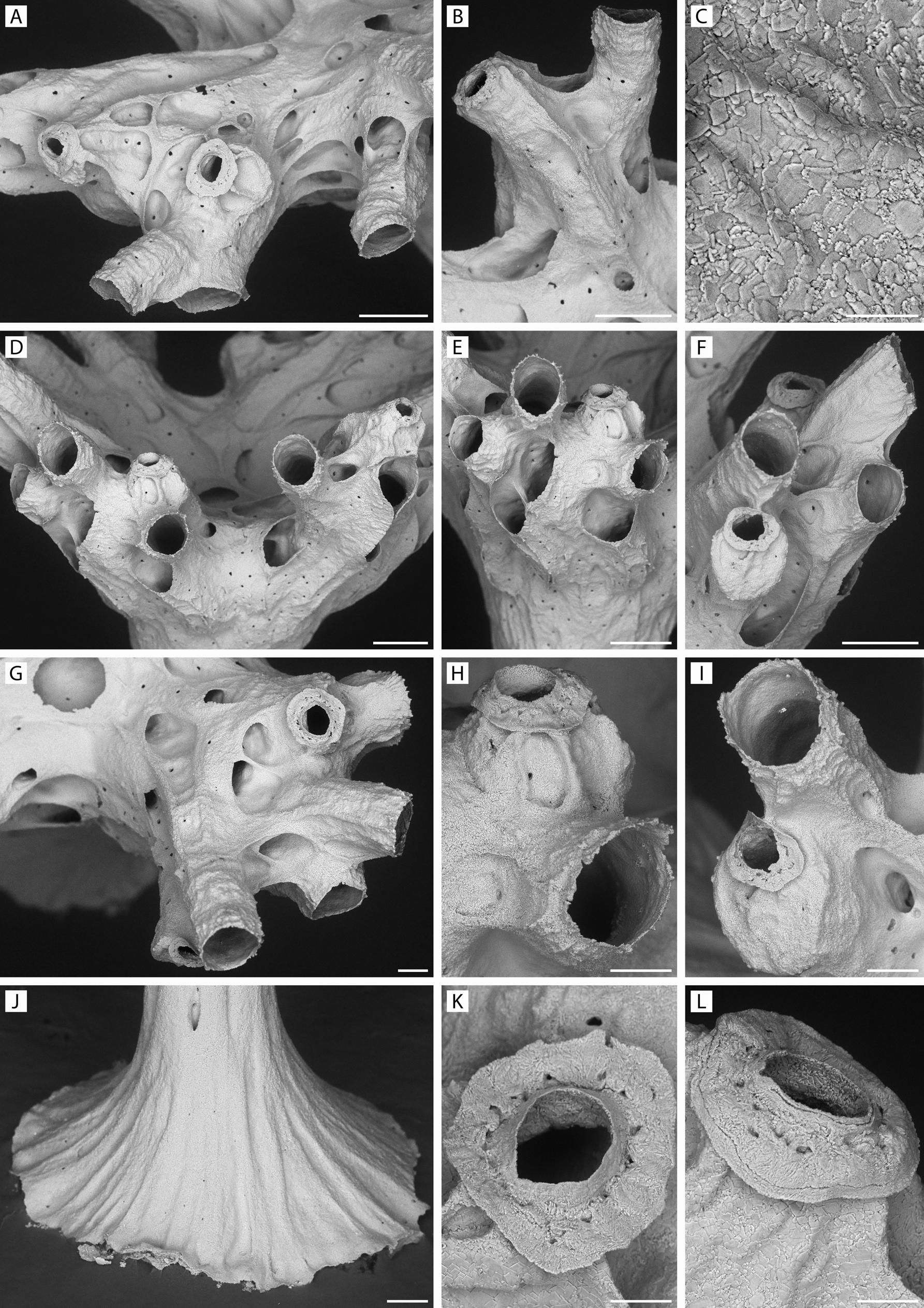
FIGURE 40. Anyuta anastema n. gen., n. sp. Holotype, ZIRAS 1/50718. A, part of fascicle with autozooidal peristomes and two dimorphic peristomes with flanges; note large shallow alveoli on sides of peristomes; B, two peristomes, left one dimorphic; C, skeletal microstructure of part of peristome in L; D–G, subfascicles with both autozooidal and flanged dimorphic peristomes; H, close-up of squat flanged dimorphic peristome in E; note small alveoli on flanks of peristome; I, close-up of lower dimorphic peristome in F, showing it to be derived from an autozooidal peristome by its partial closure; J, base of column; K, L, close-ups of perforated flanges of dimorphic peristomes. Scale bars: A, B, D, F, 250 µm; G–J, 100 µm; K, L, 50 µm; C, 25 µm.

FIGURE 41. Anyuta anastema n. gen., n. sp. Progressive stages of development of young colonies. A–D, specimen YMG4–14, Stn 360, three-zooid stage; E–H, specimen YMG4–14, Stn 338, five–six-zooid stage; I–L, specimen YMG4–13, Stn 281, and M–P, specimen YMG4–06, Stn 71, showing later stages in which trabecular walls define spaces destined to become either autozooids or alveoli. Scale bars: 200 µm.
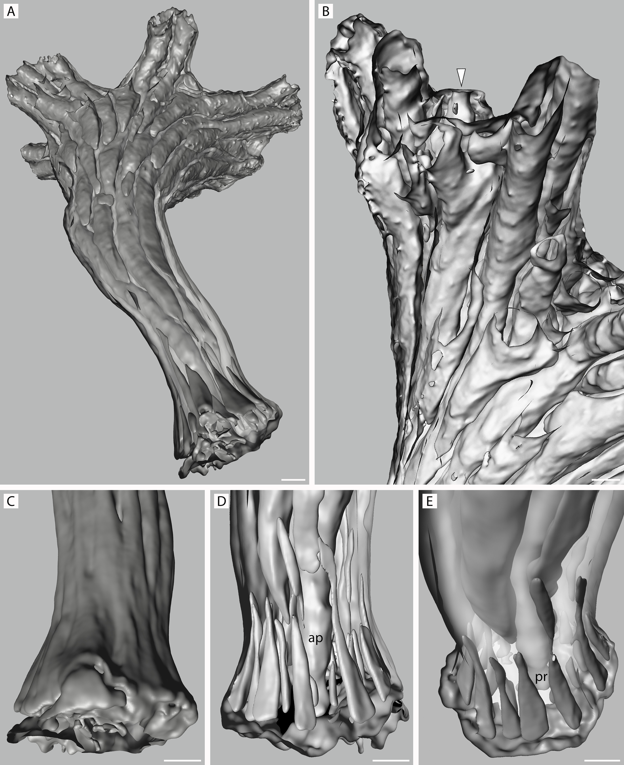
FIGURE 53. Anyuta anastema n. gen., n. sp. Micro-CT scans of paratype, NIWA 127725, as surface (C) and back-face isosurface renders showing colony interiors. A, view of colony from below; note absence of kenozooids from most of column; B, lateral abfrontal view of two small fascicles with entrance to dimorphic zooid indicated by arrowhead; note relative paucity of communication pores; C–E, exterior and interior views of colony base, showing lateral (C, D) and frontolateral (E) profiles with palisade of elongate-triangular kenozooidal chambers surrounding ancestrular protoecium (pr) and proximal part of its peristome (ap). Scale bars: 100 µm.
IMAGES
Gallery(5)
Occurrences with images
CITATIONS
References(1)
Grischenko, A. V.; Gordon, D. P.; Melnik, V. P. (2018). Bryozoa (Cyclostomata and Ctenostomata) from polymetallic nodules in the Russian exploration area, Clarion–Clipperton Fracture Zone, eastern Pacific Ocean—taxon novelty and implications of mining. <em>Zootaxa.</em> 4484(1): 1.