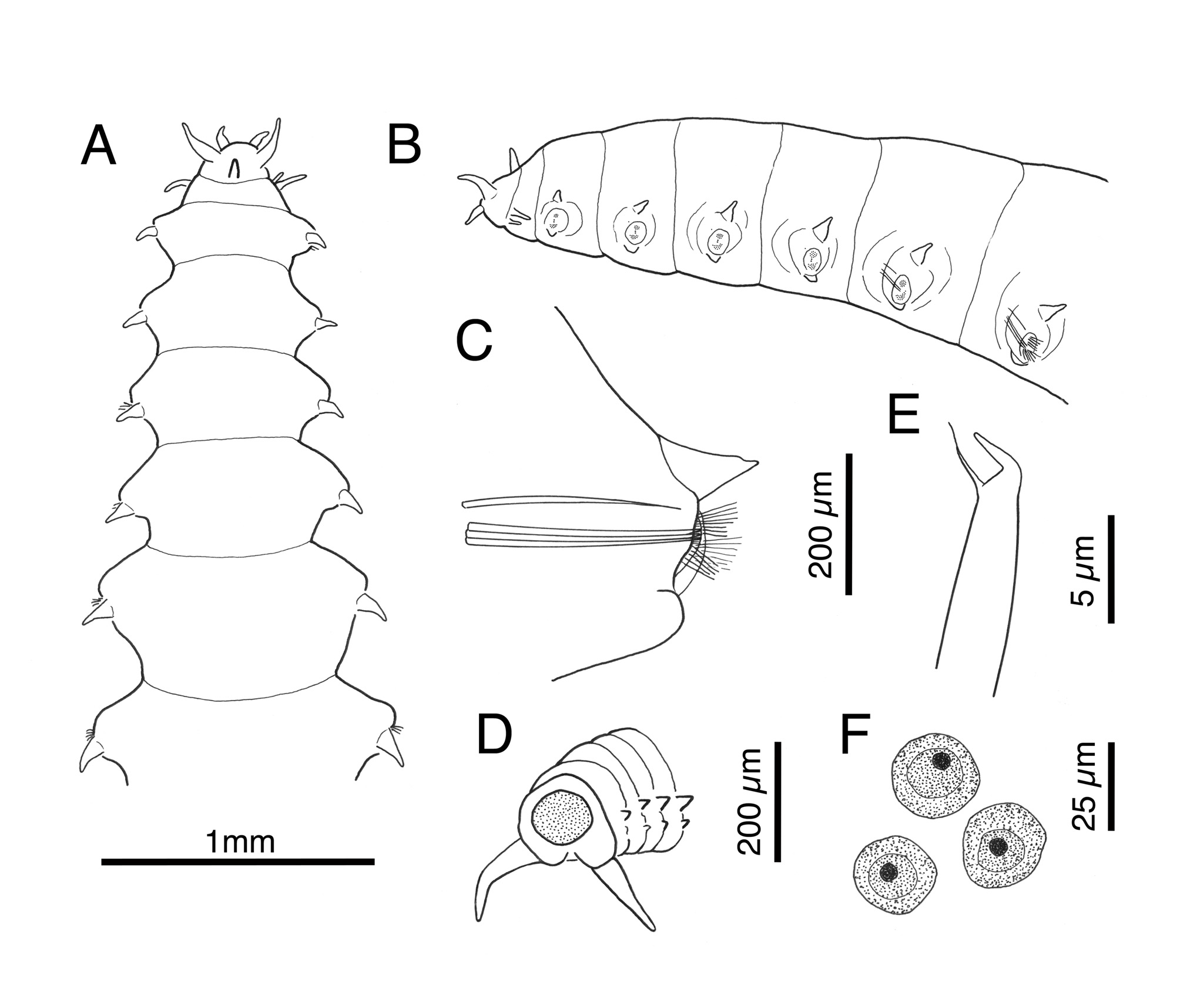
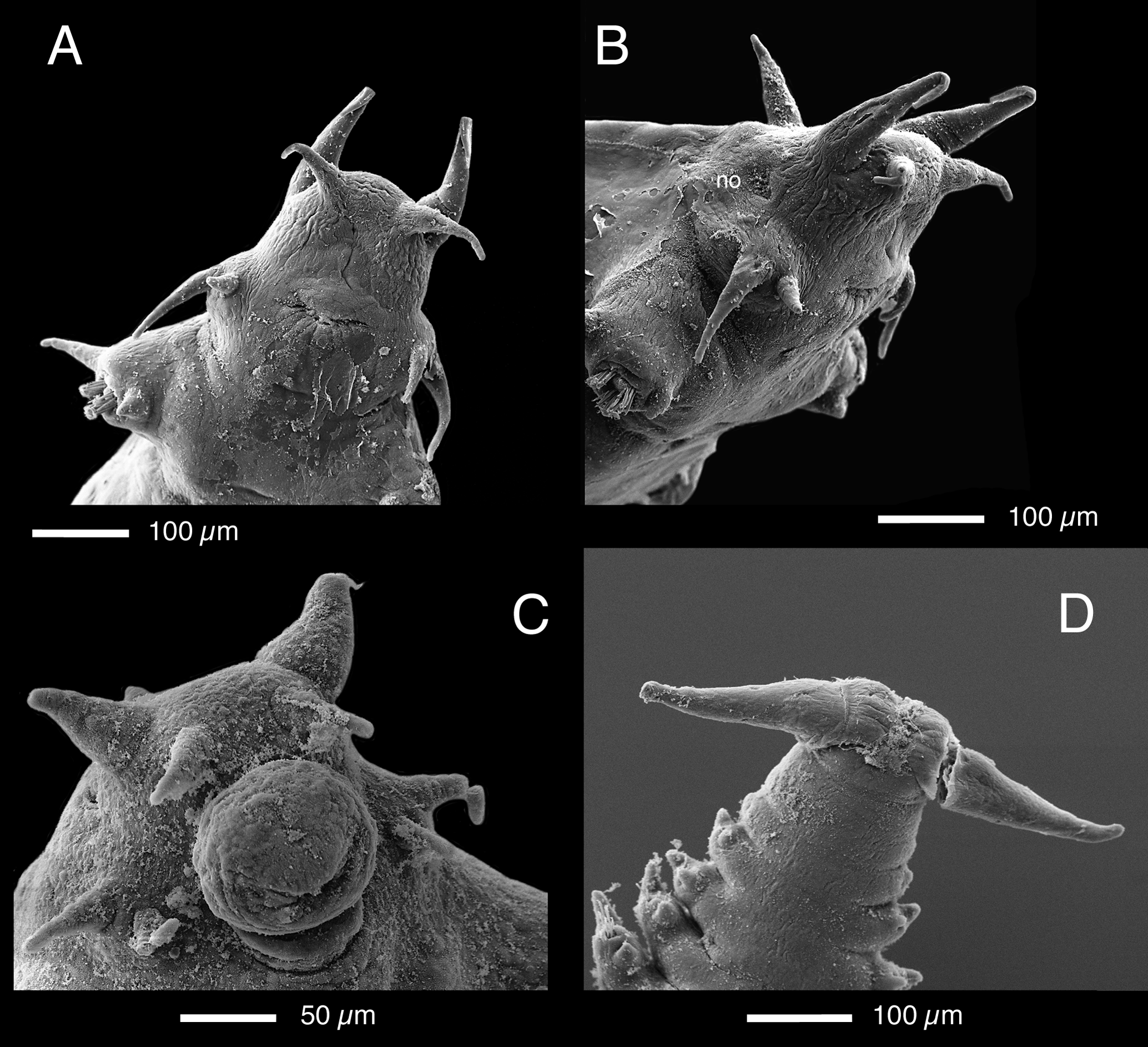
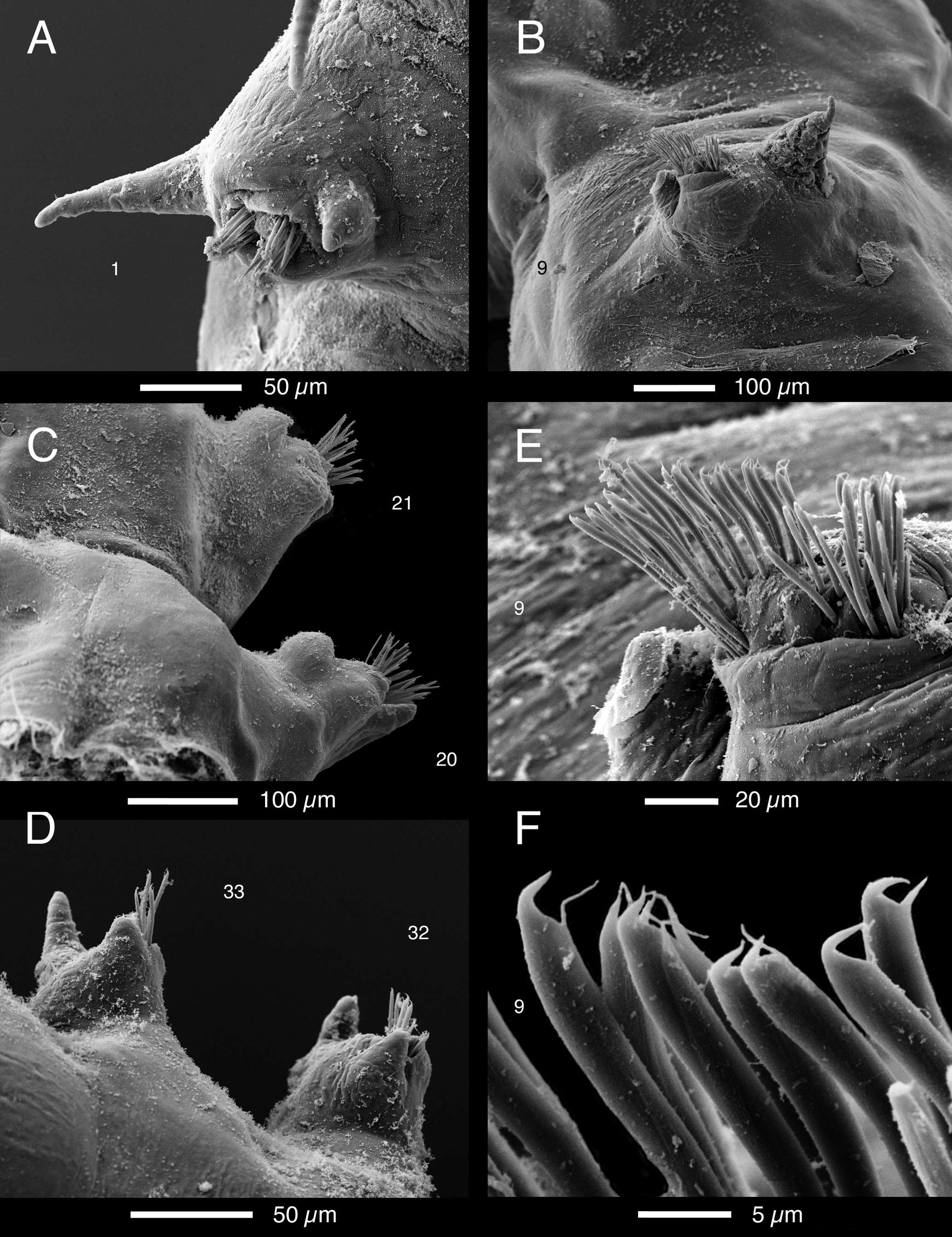
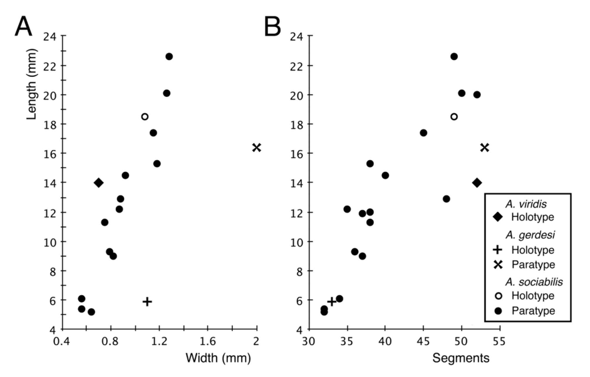
Description. Holotype entire, maximum width 1.08 mm (including parapodia), 18.5 mm long for prostomium and 48 segments. Paratypes ranging from 0.56 – 1.28 mm wide, 5.2 – 22.6 mm long, and 32 – 52 segments. Body long and slender, dorsally arched and ventrally flat, lacking ventral groove. Head and anterior 5 segments narrow; segments thereafter rapidly increasing in size (Fig. 1 A – B), reaching maximum in anterior third of body, rapidly decreasing in posterior region. Cuticle smooth and shiny on best preserved specimens. Pygidium lobulate, ventro-laterally with single pair of tapered pygidial cirri (Figs 1 D, 2 D). Prostomium subtrapezoidal, with rounded anterior margin, lacking eyes (Fig. 1 A). Pair of antero-ventral palps, somewhat conical, basally expanded and distally tapered. Pair of antero-dorsolateral antennae, of similar shape to palps, but broader and longer (Fig. 2 A – B). Median antenna of similar size and shape to palps, arising mid-dorsally near posterior margin of prostomium (Fig. 1 A, B). Nuchal organs elliptical ciliated grooves, positioned dorsolaterally between posterior prostomium and anterior margin of segment 1 (Fig. 2 B). Peristomium ventrally fused with prostomium and segment 1; delineation between prostomium and segment 1 only distinct dorsally and laterally. Mouth ventral, transverse, slit-like (Fig. 2 A – B). Two pairs of tapered tentacular cirri on segment 1, superior cirri 1.5 to 2 times as long as inferiors; parapodia and chaetae lacking. Pharynx muscular, axial, short and narrow, occupying anterior three segments; proboscis short and bulbous when extruded (Fig. 2 C), jaws lacking. Gut tubular, with markedly wider lumen from anterior to middle of chaetiger 3. Gut caecae from chaetiger 3 as pair of small lateral pouches; caecae increase in size over following 2 – 3 chaetigers, extending into parapodia. By chaetiger 6 or 7, distally tapered caecae reach into posterior parts of parapodia, just below dorsal cirri; caecae decrease in size in posterior half to third of body. Gut caecae revealed through methyl green staining; caecae appearing as opaque white unstained masses either side, extending laterally into the parapodia. Parapodia simple, short cuff-like, lacking any marked pre- or post-chaetal structures. Dorsal cirri above or slightly dorso-posterior to parapodia; cirri somewhat conical with broad bases and tapering cirriform tips of variable extension (Figs 1 A – C & 3 A – B). Ventral cirri positioned below or slightly posterior to neurochaetae. Ventral cirri of anterior and posterior parapodia short, conical with bluntly pointed tips (Fig. 3 A, D). Those of median body larger, more rounded, or rounded with tiny sharply pointed tips (Figs 1 C, 3 C). Anterior parapodia supported by two parallel, robust and distally pointed neuroaciculae; increasing to three aciculae by about chaetiger 6 or 7. Single notoaciculae with sharply tapering tips arise separate from neuroaciculae (Fig. 1 C). Each notoacicula directed forward into anterior part of parapod, tip ending anterior to and level with uppermost neurochaetae, well in advance of dorsal cirrus (Fig. 1 B, C); in slide preparations notoacicula may seem closer to dorsal cirrus than reality. Neurochaetae numerous in anterior and mid body region, disposed in two bundles; superior bundle compact, inferior bundle broader with chaetae extending more dorsally around acicular lobe on posterior side (Figs 1 B, 3 E). Number of chaetae similar in each bundle, usually slightly more in inferior bundle; two bundles together totalling about 30 – 35 chaetae in anterior parapodia, but increasing to 45 – 50 in parapodia of largest segments. Separation between superior and inferior bundles disappears in posterior parapodia, and number of chaetae decreases to less than five or six on posteriormost segments. Posteriormost 4 – 6 segments of smallest specimens have rudimentary parapodia and no chaetae. Neurochaetae slender, shafts smooth, each with strongly bent distal tooth and longer slender and delicate secondary spine, space between tooth and spine angular (Figs 1 E, 3 F); primary teeth all pointing dorsally (Fig. 3 E). Reproduction. Largest specimens, longer than about 15 mm clearly female with small (ca. 25 µm diameter) oocytes in parapodia and loose in coelom (Fig. 1 F). Several specimens between approximately 11 and 15 mm appear to be immature females. Sex of smaller specimens not determined. Colour. Preserved specimens white. Colour of live animals not known. Methyl Green staining. Glandular staining variable, most pronounced on the larger specimens. Band of speckles occurring behind the mouth and behind the ventral tentacular cirri. Ventrally, anterior 9 or 10 chaetigers with two widely separated, and interrupted, parallel lines of stain. These lines linked in posterior part of each chaetiger by broad transverse band, producing repetitive H-pattern of stain on anterior venter. The transverse band disappears on following chaetigers, and only two broken parallel lines remain. Other staining associated with the parapodia. Ventral staining patch present at base, as vertical line on body just anterior to each parapod, and as vertical patch on its anterior face. Similar, but more sparse speckled areas on posterior face of parapod. Sparse speckles of stain evident on basal part of dorsal cirri and on body dorsal to parapodia; mid-dorsal region unstained. Pygidial lobes surrounding anus stained. Morphometrics. The morphological attributes measured (width, length, number of segments) for A. sociabilis sp. nov. were strongly correlated: width and length (Fig. 4 A), r = 0.957, p <0.0001, n = 14; segments and length (Fig. 4 B), r = 0.894, p <0.0001, n = 17; segments and width (not figured), r = 0.806, p = 0.0002, n = 14.
Antonbruunia sociabilis sp. nov. (Annelida: Antonbruunidae) associated with the chemosynthetic deep-sea bivalve Thyasira scotiae Oliver & Drewery, 2014, and a re-examination of the systematic affinities of Antonbruunidae